Operetta CLS (Revvity)
High-Content Screening and Imaging
Information-rich measurements of biology in intact cells
Many important drug discovery questions cannot be answered by measuring a single biochemical endpoint. The effects of a compound may depend on cellular signaling, protein localization, receptor trafficking, cell morphology, proliferation, toxicity, or other phenotypes that emerge only in intact cells.
High-content screening combines automated fluorescence microscopy with quantitative image analysis to measure these complex biological responses. Instead of recording a single value from each well, high-content assays can capture multiple features simultaneously and analyze them at the level of individual cells and subcellular structures.
The Fisher Drug Discovery Resource Center uses high-content imaging to develop and perform cellular assays in which microscopy provides a quantitative biological readout. Our current platform is the PerkinElmer Operetta, a high-throughput, multichannel fluorescence imaging system with confocal capabilities and automated image analysis.
From Images to Quantitative Biology
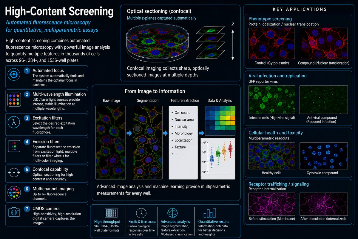
High-content screening is more than automated microscopy. The objective is to convert images into quantitative measurements that can be used for biological interpretation and compound screening.
Depending on the assay, image analysis can identify individual cells and quantify features such as cell number, nuclear morphology, fluorescence intensity, subcellular localization, cellular area, or other phenotypic characteristics.
This allows investigators to move from a qualitative observation—“the cells look different”—to quantitative measurements that can be evaluated across hundreds or thousands of experimental conditions.
Because several measurements can be collected from the same cells, high-content imaging can provide substantially more biological information than a conventional single-endpoint plate-reader assay.
Multiparametric Cellular Assays
One of the strengths of high-content imaging is the ability to measure several aspects of cellular health or phenotype simultaneously.
In one of our published studies, we developed a live-cell high-content assay called ImageTOX that measured cell number, nuclear area, nuclear intensity, and nuclear membrane permeability simultaneously. The assay was validated in a 12,668-compound screen and compared directly with a conventional ATP-based cytotoxicity assay.
The imaging assay identified compounds affecting cellular structure that were not necessarily apparent from a single metabolic endpoint. It also provided concentration-response measurements and allowed the same population of living cells to be followed over time, revealing differences in the kinetics of compound-induced cellular effects.
Importantly, the multiparametric measurements could distinguish different types of cellular responses. For example, changes in cell number could occur without corresponding changes in nuclear morphology or membrane permeability, providing information that would be difficult to obtain from a single ATP-based viability measurement.
The study also demonstrated another practical advantage of imaging-based screening: image inspection can reveal compound precipitation, fluorescent compounds, and other visual artifacts that may complicate interpretation of conventional screening assays.
Phenotypic Screening
High-content imaging is particularly powerful when the desired screening endpoint is a cellular phenotype rather than a single predefined biochemical activity.
For example, in a collaboration with the Hudspeth laboratory, we used a high-content phenotypic screening strategy to identify small molecules that promote nuclear translocation of the transcriptional regulator Yap in confluent MCF10A cells.
The assay was designed around a biological phenotype: under conditions of high cell density, Yap is normally excluded from the nucleus. Compounds that altered this behavior could therefore be identified by measuring the fraction of cells displaying nuclear Yap.
The assay also incorporated a cellular health counter-screen based on cell number, allowing compounds that simply reduced cell survival to be eliminated. Six compounds met the criteria established for the primary screen, leading to further characterization of the compounds and ultimately to the identification of TRULI, a potent inhibitor of Lats1 and Lats2.
This example illustrates the distinctive value of high-content screening: the primary assay did not simply ask whether a compound was active. It asked whether the compound produced a specific spatial and biological change within the cell.
Viral Infection and Replication
High-content imaging can also be used to measure viral infection and replication in intact cells. Viruses can be engineered to express fluorescent reporters such as GFP or RFP during their lifecycle, allowing infection and viral replication to be monitored directly by automated fluorescence imaging.
This approach provides a quantitative cellular readout of antiviral activity while simultaneously measuring effects on cellular health. Image analysis can quantify the number or intensity of fluorescently infected cells while also measuring cell number, morphology, or other indicators of cytotoxicity.
This is particularly useful for distinguishing antiviral activity from nonspecific cellular toxicity. A compound that reduces viral reporter signal but also causes substantial loss of cells may have a very different interpretation from a compound that suppresses viral infection while leaving the cells healthy.
The same general approach can be adapted to different stages of the viral lifecycle, depending on the reporter and viral system used. Replicons and other engineered reporter systems can provide additional experimental strategies for studying viral replication and pathway-specific effects.
SARS-CoV-2 and Antiviral Drug Discovery
The DDRC has applied image-based cellular assays to antiviral drug discovery, including our work on small-molecule inhibitors of the SARS-CoV-2 NSP14 RNA cap methyltransferase.
In this work, fluorescent reporter-based cellular assays provided a way to determine whether compounds identified through biochemical screening produced the expected antiviral effect in cells. Image-based measurements allowed viral infection to be evaluated together with cellular health, providing an important cellular bridge between molecular target inhibition and antiviral activity.
This illustrates an important role for high-content imaging in drug discovery: connecting inhibition of a molecular target with an observable effect on viral biology in intact cells, while simultaneously monitoring cellular toxicity.
What Can High-Content Imaging Measure?
The appropriate readout depends upon the biological question. Potential applications include:
- Protein localization and nuclear translocation
- Receptor internalization and trafficking
- Cell proliferation and cell number
- Cell death and cytotoxicity
- Cell morphology
- Nuclear morphology
- Protein aggregation
- Organelle morphology and function
- Cellular differentiation
- Intracellular signaling
- Phenotypic responses to small molecules
- Viral infection and replication
- Fluorescent reporter-based viral assays
- Viral protein expression and localization
- Simultaneous measurement of antiviral activity and cellular toxicity
Receptor Biology and Cellular Signaling
High-content imaging is particularly useful for biological systems in which signaling produces changes in protein localization, receptor trafficking, or cellular morphology.
For example, receptor internalization can provide a complementary readout for GPCR projects alongside calcium flux, cAMP, IP1, β-arrestin, or reporter-gene assays.
Similarly, nuclear translocation of signaling proteins can provide a direct cellular readout of pathway activation. These measurements can sometimes provide mechanistic information that is not accessible from a downstream bulk biochemical endpoint.
Assay Development
Developing a successful high-content screening assay requires more than obtaining a good fluorescent image. The biological phenotype must be robust, reproducible, and quantifiable, and the image-analysis workflow must reliably distinguish meaningful biological responses from technical variation.
Assay development may include optimization of:
- Cell type and culture conditions
- Cell density and plating conditions
- Fluorescent probes and antibodies
- Staining conditions
- Incubation time
- Image acquisition parameters
- Confocal imaging conditions
- Cell and subcellular segmentation
- Image-derived quantitative features
- Controls and normalization
- Plate format and assay miniaturization
An important part of development is determining which image-derived features actually answer the biological question. More measurements are not necessarily better. The goal is to identify robust, biologically meaningful endpoints while minimizing unnecessary complexity in the screening and analysis workflow.
Live-Cell Imaging and Kinetics
When an assay can be performed using living cells, high-content imaging can provide an additional dimension of information: time.
Rather than measuring the final state of a cell population at a single endpoint, investigators can follow changes in the same population over time. This can reveal differences in the kinetics of compound activity and help distinguish early cellular responses from later consequences of treatment.
Our ImageTOX work demonstrated this principle by following multiple cellular health parameters over extended treatment periods. Compounds that produced similar levels of toxicity at a late time point could have substantially different temporal profiles.
Time-dependent measurements can therefore provide information about mechanism that is unavailable from conventional single-endpoint assays.
High Content as Part of a Screening Cascade
High-content imaging is rarely an isolated technology. It can be used as a primary screening method, as a secondary assay, or as an orthogonal method for understanding the biological consequences of compound activity.
A project might begin with a biochemical assay and use high-content imaging to determine what active compounds do in intact cells. Conversely, a phenotypic high-content screen may identify compounds whose molecular mechanism is subsequently investigated using biochemical and biophysical assays.
In antiviral discovery, a project may begin with a biochemical assay against a viral enzyme and progress to a cellular assay that measures viral infection or replication. Fluorescent reporter systems can provide a direct cellular readout of whether inhibition of a molecular target produces the expected effect on viral biology.
For this reason, high-content imaging fits naturally into a broader discovery cascade:
Biological question → cellular phenotype → high-content screening → hit confirmation → mechanism → biochemical and biophysical characterization
Why Use High-Content Imaging?
- Information-rich: multiple biological measurements can be obtained from the same experiment.
- Single-cell resolution: heterogeneous responses can be distinguished rather than averaged across an entire well.
- Spatial information: protein localization, trafficking, and morphology can be measured directly.
- Phenotypic: compounds can be identified based on cellular responses without requiring a fully defined molecular mechanism at the outset.
- Multiparametric: several features can be combined to characterize compound responses.
- Kinetic: live-cell assays can sometimes follow biological responses over time.
- Artifact detection: images can reveal precipitation, fluorescence, morphology changes, and other effects that may complicate interpretation of screening data.
- Cellular mechanism: imaging can connect molecular target activity with changes in intact cells.
Considerations and Limitations
High-content imaging generates substantially more information—and therefore more data—than a conventional plate-reader assay. The quality of the results depends on cell health, staining consistency, image quality, segmentation, feature selection, and the robustness of the image-analysis workflow.
Image analysis can also introduce additional sources of variability. For this reason, assay development should include careful evaluation of controls, reproducibility, signal window, image quality, and the biological relevance of the selected features.
High-content imaging is therefore not necessarily the best solution for every cellular assay. When a simple biochemical or plate-reader endpoint adequately answers the biological question, a simpler assay may provide greater throughput, lower cost, and easier analysis.
DDRC Perspective
High-content imaging is most valuable when the biology itself is complex.
Our experience ranges from measuring cellular toxicity and morphology to identifying specific changes in protein localization and signaling, and to measuring viral infection and replication in cellular systems.
The goal is not simply to acquire more images. It is to identify the biological phenotype that matters, develop a reliable way to measure it, and connect that measurement to the molecular mechanism being investigated.
This makes high-content imaging particularly powerful in drug discovery: it can provide the experimental bridge between a molecular hypothesis and what actually happens inside a living cell.
Our published work illustrates both sides of this approach: developing information-rich cellular assays for large compound screens and applying phenotypic imaging to discover compounds that alter a specific biological pathway.
At the DDRC, high-content imaging is a tool for turning complex cellular biology into quantitative information that can drive drug discovery.
Selected DDRC Publications
Chiaravalli J, Glickman JF. A High Content Live Cell Viability Assay and its Validation on a Diverse 12K Compound Screen.
Small-molecule inhibition of Lats kinases promotes Yap-dependent proliferation in postmitotic mammalian tissues. Collaboration with the Hudspeth laboratory.