Chirascan V100 Circular Dichroism Spectrometer
Circular dichroism spectroscopy for characterization of protein secondary structure, conformational changes, and thermal stability.
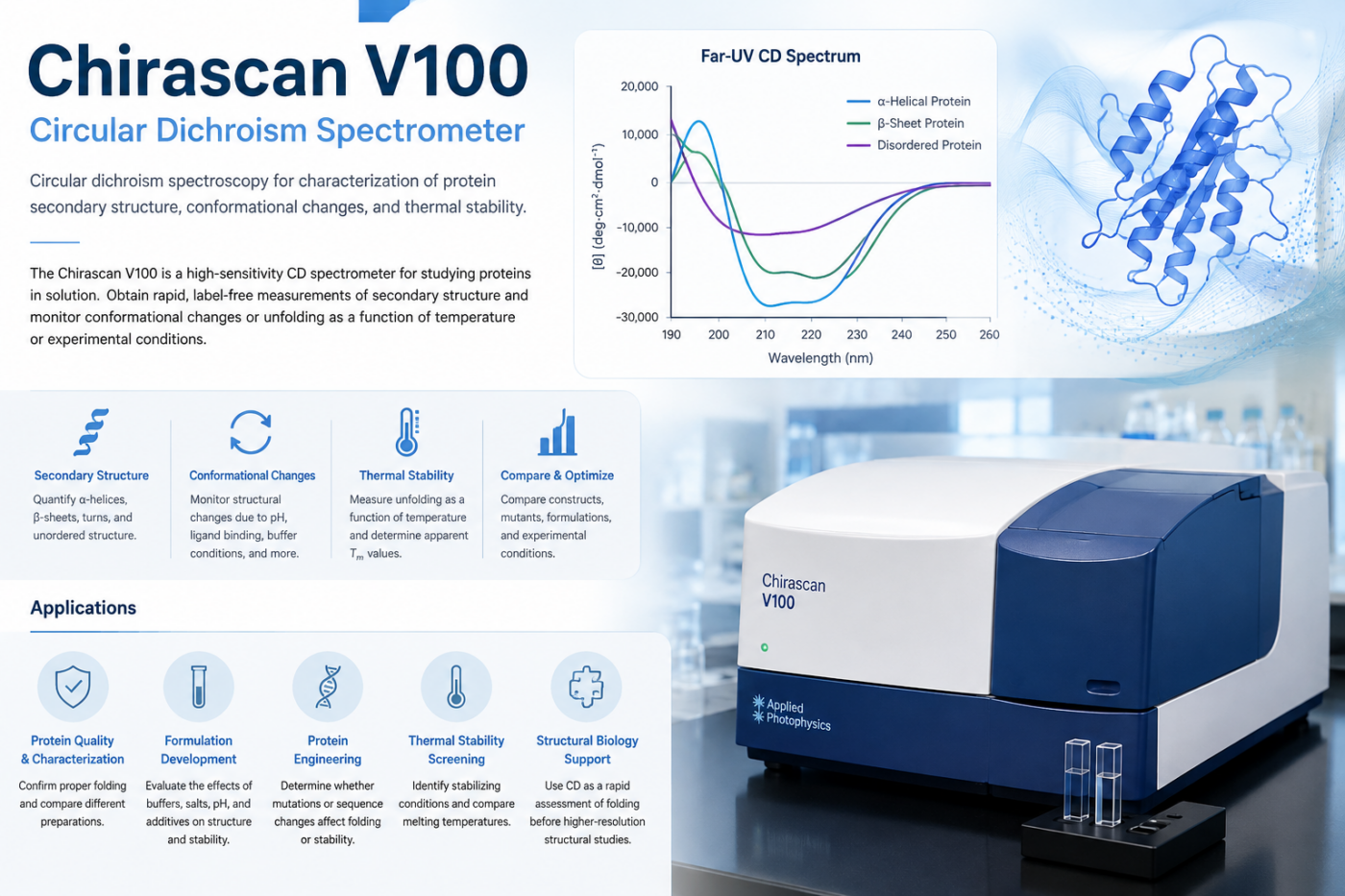
The Chirascan V100 is a high-sensitivity circular dichroism (CD) spectrometer for studying protein structure and stability in solution. CD provides a rapid, label-free way to assess protein secondary structure, monitor structural changes, and measure thermal unfolding.
At the DDRC, CD spectroscopy can be used as a standalone characterization method or as part of a broader biophysical characterization workflow.
What Can CD Spectroscopy Tell You?
Circular dichroism is particularly useful when you need to determine whether a protein is properly folded, whether its structure changes under different conditions, or how stable it is.
Protein Secondary Structure
Far-UV CD spectra provide information about the secondary structure of proteins, including the relative contributions of:
- α-helices
- β-sheets
- Turns
- Disordered or unstructured regions
CD can therefore be used to compare proteins, constructs, mutants, or protein preparations and to determine whether a purification or formulation change has altered the protein’s structural state.
Conformational Changes
CD can monitor structural changes caused by changes in:
- Temperature
- pH
- Buffer composition
- Ionic strength
- Ligand or cofactor binding
- Mutations
- Protein concentration
- Formulation conditions
Comparing spectra before and after an experimental perturbation can provide a rapid indication of whether the protein undergoes a significant conformational change.
Thermal Stability
Temperature-dependent CD measurements can be used to follow protein unfolding and determine an apparent melting temperature (Tm).
Thermal unfolding measurements are useful for:
- Comparing protein constructs
- Screening buffer and formulation conditions
- Evaluating stabilizing compounds
- Assessing the effects of mutations
- Characterizing protein preparations
- Supporting protein developability studies
Why Use CD?
CD is a relatively simple and rapid technique that requires no fluorescent or radioactive labeling. Because measurements are made directly on proteins in solution, CD can provide a useful orthogonal measurement of structural integrity alongside other biophysical assays.
A typical workflow might include:
Protein preparation → CD spectrum → secondary-structure assessment → thermal unfolding → comparison of conditions
CD can also complement techniques such as DLS, thermal-shift measurements, SPR, MST, and ITC, providing structural information that is not directly obtained from binding or size measurements.
Applications
Protein Quality and Characterization
Confirm that a purified protein has the expected structural characteristics and compare different preparations or constructs.
Formulation Development
Evaluate the effects of buffers, pH, salts, additives, and other formulation components on protein structure and stability.
Protein Engineering
Compare mutants and engineered constructs to determine whether sequence changes affect folding or stability.
Thermal Stability Screening
Measure and compare protein melting behavior under different experimental conditions.
Structural Biology Support
Use CD as a rapid solution-phase assessment of protein folding and secondary structure before committing to more intensive structural studies.
What We Can Measure
Far-UV CD spectra
Characterization of protein secondary structure and changes in structural composition.
Thermal unfolding profiles
Monitoring the loss of secondary structure as temperature increases and determining apparent Tm values.
Condition-dependent structural changes
Comparison of protein structure across buffers, pH values, ligands, salts, and other experimental conditions.
Comparative protein characterization
Evaluation of mutants, constructs, batches, and preparation conditions.
Working With the DDRC
The DDRC can help with experimental design, sample preparation considerations, measurement, and data interpretation. CD experiments can be designed around a specific scientific question rather than simply generating a spectrum.
If you are trying to determine whether a protein is properly folded, identify conditions that stabilize it, compare protein variants, or understand a conformational change, we can help determine whether CD is an appropriate approach and how it can be combined with complementary biophysical measurements.
Related Capabilities
Dynamic Light Scattering (DLS)
Assess protein size, aggregation, and sample homogeneity.
Thermal Stability Analysis
Compare protein stability across conditions and identify stabilizing environments.
Surface Plasmon Resonance (SPR)
Measure protein–protein and protein–small-molecule interactions.
Microscale Thermophoresis (MST)
Measure molecular interactions using small quantities of sample.
Isothermal Titration Calorimetry (ITC)
Characterize binding affinity and thermodynamic parameters.
Instrument
Chirascan V100 Circular Dichroism Spectrometer
Applied Photophysics