Glial cells, not neurons, lead the way in brain assembly
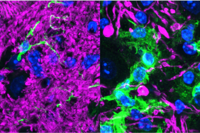
Brain formation in worms goes awry when glial cell signaling is disrupted (right).
As the very first neurons come together to form the brain, they need pointers to end up in the right places. Where do these directions come from?
Rockefeller scientists have discovered that they originate from an unlikely source, revealing that the cells directing the very first steps of brain assembly are not other neurons, as scientists have long assumed, but so-called glial cells.
“While glial cells are abundant in the brain, their functions are much less understood,” says Shai Shaham, Richard E. Salomon Family Professor. “We’ve shown that in the roundworm C. elegans, glial cells play a pivotal role, coaxing neurons onto a specific path so that proper brain assembly can ensue.”
Shaham and his team also found that these worm glial cells are remarkably similar to their vertebrate counterparts.
To peek inside the earliest stages of brain development, scientists must observe and analyze embryos, which are notoriously tricky to study. But research associate Georgia Rapti, the lead author of a recent Nature Neuroscience report describing this work, was able to use C. elegans as a model to tease apart how brain assembly begins.
“We found that glial cells grow radial processes, or extensions, marking where the first axons must go, and they release multiple signals to guide neurons onto the correct path,” says Rapti. “Our experiments show that when glial cells or their signals are compromised, the worm’s brain is severely disrupted—more than 60 percent of its neuronal extensions fail to enter the brain as they normally would.”
Rapti and Shaham were also able to show that once glial cells have set the stage for brain assembly, a special group of 10 neurons, called pioneer neurons, are the first to follow suit. Pioneer neurons had previously been documented in other species, but their identities, molecular functions, and growth properties were generally unknown.
The researchers discovered a molecular signature for pioneer neurons, and showed that these cells can act with glial cells to recruit follower neurons. They were also able to uncover previously hidden molecular pathways that glial cells and pioneer neurons use to attract the next set of neurons into the brain.
While C. elegans may not seem to have a lot in common with humans and other vertebrates, their brain assembly processes resemble ours at the molecular level. For example, Rapti and Shaham note recent reports revealing that the assembly of the mouse spinal cord relies on a signaling molecule called Netrin, which is produced by radial glial cells. The researchers found that Netrin functions similarly in the worm—glial cells produce this molecule, which guides pioneer neurons to form the C. elegans brain.
“The glial cells driving brain assembly in the worm look like—and function similarly to—those in the vertebrate spinal cord,” says Shaham. “We think of glial cells as the Pied Piper for nervous system assembly across animals, and the Piper’s pipe appears to be the same from worms to mammals.”