The secret life of chromosomes
 COPY LINK
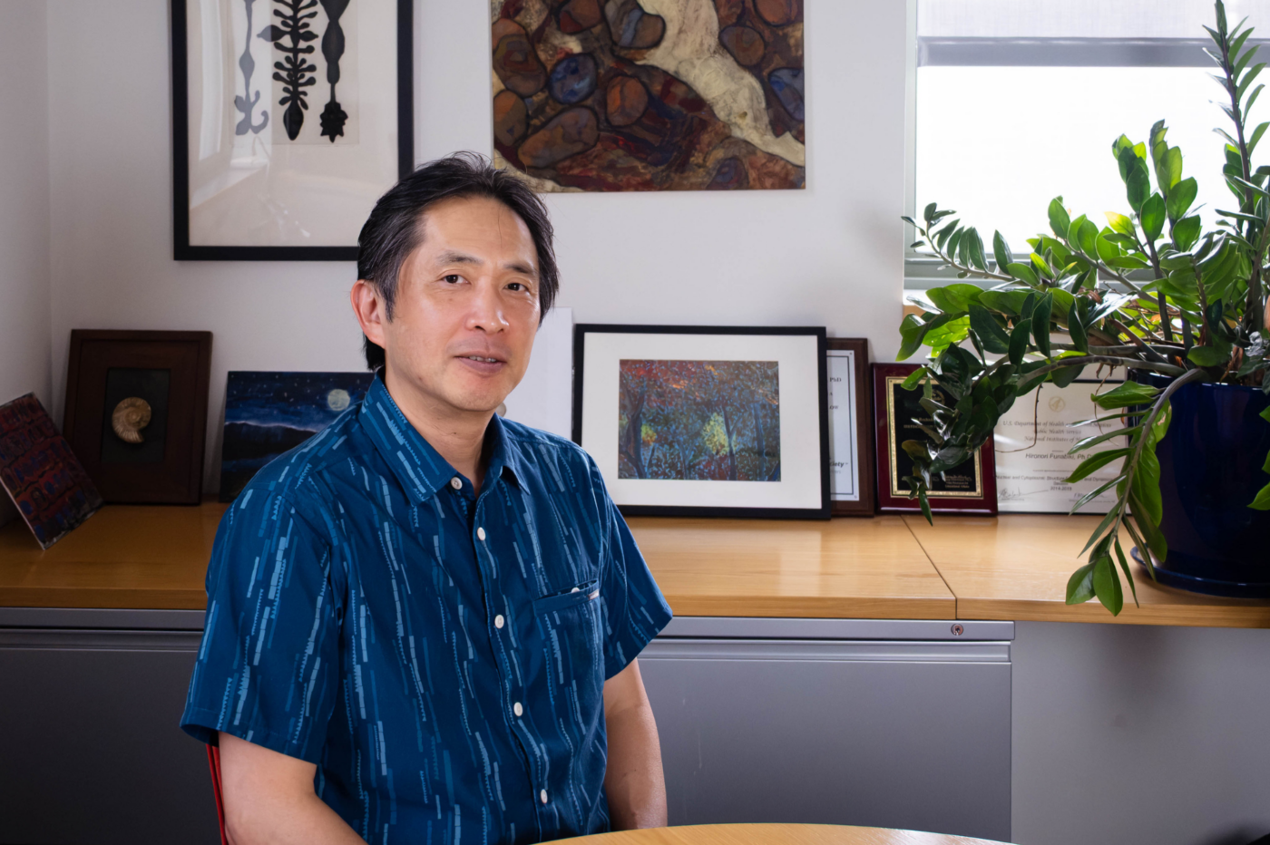
Hironori Funabiki studies how the chromosomes containing all our DNA are copied and maintained. (Credit: Lori Chertoff)
Trillions of cells in your body each carry 46 chromosomes—tightly wound packages of DNA that contain all your genes and the instructions for using them. These vital structures must be constantly maintained, accurately copied, and distributed every time a cell divides. If a dividing cell sends 45 chromosomes to one of its daughter cells and 47 to the other, the resulting cells may die or trigger disease. A single error or piece of unrepaired damage in a chromosome can lead to cancer, immune disease, or accelerated aging.
For nearly 150 years, scientists have been able to peer through a microscope and watch a cell as it divides into two, a process called mitosis. Yet the molecular machinery that controls the precise dance of chromosomes throughout this process is still not fully understood.
Hironori Funabiki, professor and head of the Laboratory of Chromosome and Cell Biology, is trying to change that. His lab investigates how chromosomes are copied and doled out during cell division, how their chemical markings are kept intact, and how cells recognize their own chromosomes as different from the DNA of invading viruses.
We spoke to Funabiki about what drew him to chromosomes, what his research has revealed, and what implications his work has for medicine.
How did you end up focusing your lab on chromosomes?
I grew up in Kyoto, Japan and had always been drawn to philosophy. As an undergraduate, I began to become interested in biology because I realized that to understand the core of who we all are and what consciousness means, you need to understand how the brain works. But at the time in Japan, there weren’t a lot of opportunities to study these kinds of things, so I ended up in a lab studying mitosis and chromosome segregation. Through that work, I realized that studying how chromosomes are copied and distributed is actually one of the most fundamental and profound questions in biology.
My mentor, Mitsuhiro Yanagida, showed me that this question of how a cell divides its chromosomes touches on the deepest questions about life. How is biological information preserved across generations? How does a cell know what belongs to it and what doesn’t? How does the identity of a cell persist over time?
Today my lab focuses on what I call “the three Is of chromosome biology.” Inheritance, or how chromosomes and their encoded information are accurately transmitted through mitosis; integrity, how long and fragile genomic DNAs are folded into physically robust chromosomes with a variety of functional features; and identity, how cells distinguish their own chromosomes from foreign DNA.
Cell division happens constantly in every living thing. What can go wrong with chromosomes during this process?
There’s a Tolstoy quote that goes something like: happy families are all alike, but every unhappy family is unhappy in its own way. I think about chromosomes the same way. When chromosome division works correctly, it always works the same way. But there are so many ways it can go wrong.
For chromosomes to get distributed during cell division, they have to attach to spindle fibers—the molecular threads that pull the chromosomes to the two opposite ends of the cell. When that attachment goes wrong, one daughter cell ends up with too many chromosomes and the other with too few. This happens frequently in cancer.
We’ve been trying to work out how healthy cells detect and correct these errors. We’ve recently made headway in understanding how spindle fibers themselves play a role. That finding gives us a more precise picture of where the system breaks down in cancer cells.
You use frog eggs to study mitosis. Why?
Human cells need to remain intact for their chromosomes to arrange themselves into spindles and divide up. But with frog eggs, when you gently extract the internal material and then add DNA, within minutes the proteins all go to work. DNA is organized into chromosomes and spontaneously assembles the spindle, just as it would inside a living cell. The first time you see it is so exciting; it’s almost alive.
Because these components are all free-floating in a kind of gel, you can then add or remove specific proteins, change the DNA that you’re adding, and watch what happens in real time. We use magnetic beads attached to the DNA so we can pull it out of the mixture at any time and see what proteins have bound to the DNA.
We’ve also recently developed a new cryo-electron microscopy method called MagIC-cryo-EM that lets us see what’s happening during this process with near-subatomic resolution. That system is what let us study how genomic DNAs are folded into chromosomes. But because our method greatly reduced the amount of a sample needed for structural analysis of biological materials, we’ve been gratified to see that it’s now being utilized in a variety of questions in life and medical sciences.
What have you found when it comes to chromosomes and immune disease?
One of our discoveries which I’m very proud of, and came about in a very unexpected way, was identifying this protein CDCA7 related to a rare immune disease called ICF syndrome, in which patients have severely compromised immune systems.
All chromosomes contain not only the sequences of DNA letters that encode genes, but also chemical tags called methylation attached to the DNA. When DNA is copied, these patterns of methylation also have to be copied.
Using frog eggs, we discovered that CDCA7 acts as a sensor, detecting a very specific methylation pattern that appears on newly copied chromosomes. It then recruits the machinery needed to mark up the new chromosome with the correct methylation. When CDCA7 is mutated, that process fails and the methylation pattern of chromosomes changes.
This may potentially explain what goes wrong in ICF patients; immune cells can’t maintain correct methylation when carrying out the rapid cell proliferation needed to mount an immune response. It turns out to also have implications in cancer, since cancer cells appear to have difficulty maintaining DNA methylation pattern as they proliferate. In both cases, understanding the process might lead us to ways to target it with therapies.
Your lab has also found unexpected connections between chromosomes and how our bodies detect viruses. What’s going on there?
Scientists know that cells have a sensor for foreign DNA, cGAS, that triggers the immune system to respond—this is how cells detect invading viral DNA. But we’ve never fully understood why cGAS doesn’t set off the alarm against our own chromosomes.
We discovered that it has to do with how chromosomes are packaged into their three-dimensional structures. Chromosomes aren’t just balls of DNA; they’re folded into structures called nucleosomes. We showed that the nucleosomes are what is responsible for silencing cGAS. Viruses penetrate cells without any nucleosomes and don’t have that ability to shut off the sensor.
Do you think studying chromosomes is getting you closer to answering those big philosophical questions you had early in your career?
I do. I like work that can promote the benefit of humanity and if we can cure a disease, that would be enormously meaningful. But I’m still also driven by philosophy. And I think it all ties into chromosomes.
The molecules constituting your body are replaced over time. The chromosomes in your cells today carry largely the same DNA sequences you were born with, but the chemical marks on them have been copied and re-copied billions of times, accumulating small changes along the way. How do I know that I’m the same person as when I was five years old, if most of my cells are new? We tend to think of our brains—our consciousness—as the part of us that maintains our identity, but your identity lives in cells throughout your body.
I think it’s important to keep thinking about these kinds of fundamental questions to better understand the nature of life and what it means to be human.