Three-dimensional model illuminates key aspects of early development
Researchers used a 3D model based on lab-grown stem cells to simulate the moment in development when the body starts to separate into two distinct halves.
From a biological standpoint, the earliest stages of life are the most mysterious. A developing human embryo undergoes a flurry of rapid changes, and these changes are exceedingly difficult to study because they transpire within the confines of a womb.
But with new technology, it might soon be possible to fill important gaps in our understanding of early pregnancy and development. Rockefeller scientists recently used stem cells to create a 3D model of early embryonic tissues, allowing them to simulate developmental processes as they occur in time and space. The researchers hope that this tool, whose utility they recently demonstrated in a report in Nature Cell Biology, will make it possible to further elucidate the processes that guide embryonic growth, and ultimately lead to innovations that promote healthy pregnancies.
Dimensions of development
The concept of using stem cells to model early embryonic development was first developed in labs of Ali H. Brivanlou, the Robert and Harriet Heilbrunn Professor, and Eric D. Siggia, the Viola Ward Brinning and Elbert Calhoun Brinning Professor, who published their initial research on the topic in 2014. Despite making several crucial discoveries since that time, Brivanlou and Siggia knew that his system was, in some ways, limited: conventional stem cell models are two dimensional and do not take on the actual shape of an embryo, therefore prohibiting researchers from asking key questions related to its structure.
For example, scientists are highly interested in the process by which embryos attach to the uterus—a crucial first step to a successful pregnancy. And according to Mijo Simunovic, a Simons Junior Fellow in Siggia’s lab, it is practically impossible to study this complex phenomenon in a two-dimensional system.
“Attachment is inherently a 3D problem,” he says.
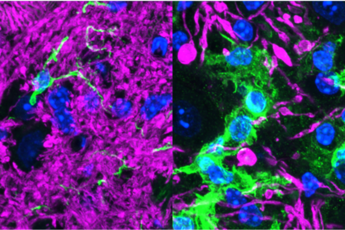
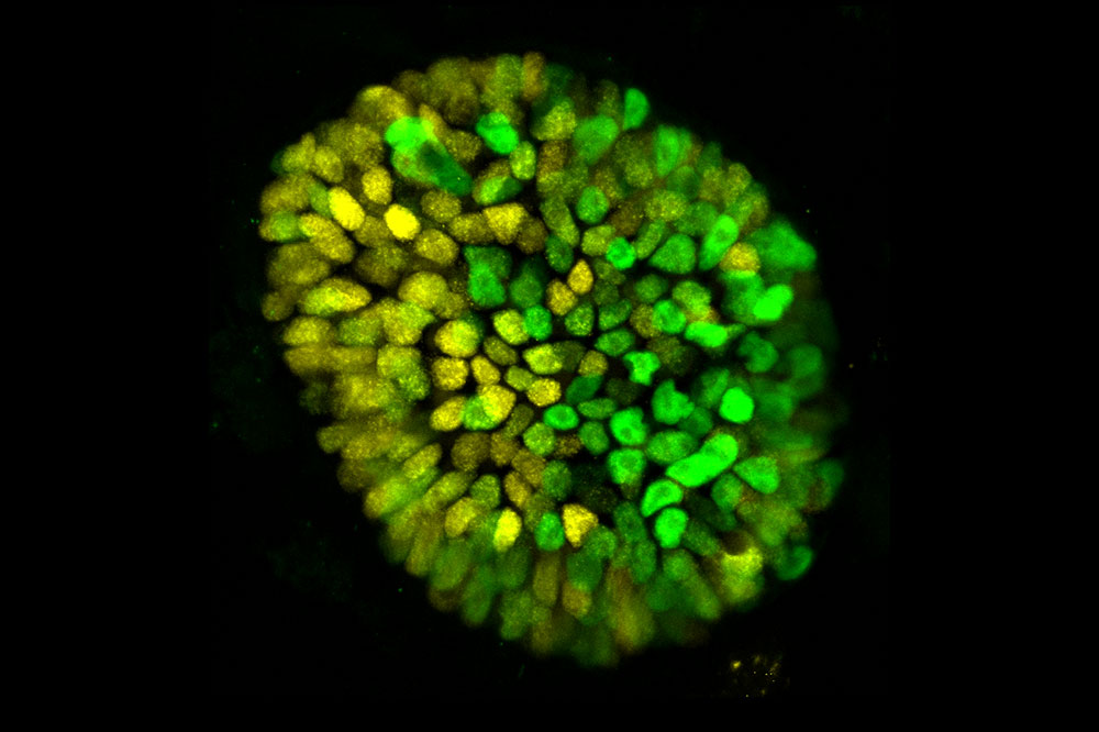
To address this issue, Brivanlou, Siggia, and Simunovic used an interdisciplinary approach to develop a 3D model simulating an approximately fourteen-day-old human embryo—the stage of development during which a key milestone of embryonic development called “gastrulation” takes place.
“We combined several techniques—bioengineering, physics, and developmental biology—to create this model,” explains Simunovic, who notes that this research would not have been possible without the unique, longstanding collaboration between the Siggia and Brivanlou labs. “We now have a 3D system that mimics not only the embryo’s genetic fingerprint, but also its shape and size.”
Of course, it is not enough to make a model that simply looks like a real embryo: it must also act like one. Accordingly, the researchers tested whether their system could simulate one of the most fundamental processes in animal development—a phenomenon known as symmetry breaking.
Breaking symmetry, making progress
In its earliest stage, an embryo comprises a symmetrical sphere of cells. Then, after about two weeks, this symmetry begins to disappear as the embryo takes on distinct features that will become various parts of the body.
“Symmetry breaking drives almost everything that happens during embryonic development,” says Simunovic. “Our heads don’t look like our feet, and that’s because, at some point, the embryo breaks into two parts, anterior and posterior.”
This breakage is, in fact, the first symmetry breaking of the body axis that takes place during human development, and it transpires just after attachment to the uterus. If the researchers could induce such a breakage in their model, they reasoned, then they would know that their system accurately emulated a real embryo—at least during this key period in developmental time.
To this end, the researchers exposed their model to chemical signals that, in pregnancy, are released by the placenta. Through a series of experiments, they found that the addition of a chemical known as BMP4 reliably prompts symmetry breaking.
“We added BMP4, and two days later one part of the three-dimensional culture became the future posterior, and the opposite part became the future anterior,” says Simunovic.
This result has implications beyond elucidating the chemistry of a particular developmental process. Now that scientists can successfully model embryonic events in 3D, extensions of this research may be used in future studies of pregnancy complications, such as unsuccessful attachment.
“About 50 to 75 percent of embryos do not attach, creating a huge bottleneck to pregnancy,” says Simunovic. “We don’t know why that is, but using this model we may be able to find out.”
This system, Brivanlou notes, could also be used to study inborn diseases. “We can create 3D embryonic models of genetic conditions, and follow the developmental process in real time,” he says. “These models can finally advance the understanding of a wide range of diseases for which we currently have no idea where and when things begin to go wrong.”