An unexpected origin for calming immune cells in the gut
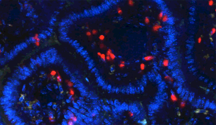
Disappearing T regs: When researchers tracked calming immune cells, known as T regs, they found these cells (red) usually stayed in the tissue below the single-cell lining (blue) of the gut, and when they did enter the lining, many did not return.
Biologically speaking, we carry the outside world within us. The food we ingest each day and the trillions of microbes that inhabit our guts pose a constant risk of infection—and all that separates us from these foreign entities is a delicate boundary made of a single layer of cells.
The immune cells that swarm about this threshold must exercise a precise balancing act. They must be vigilant, yet also tolerant of harmless substances so that they don’t cause harmful overreactions. New research at The Rockefeller University described in the June 24 issue of Science explains how a specific set of immune cells in the gut originate to help maintain this equilibrium.
Inflammation is a key element of the body’s nonspecific response to a threat, but if activated inappropriately, it can damage tissue, or lead to allergies and autoimmune disease. Throughout the body, white blood cells called regulatory T cells, or T regs, are tasked with calming the inflammatory response.
“But T regs are strangely absent in the thin lining of the gut—a place where one would expect to see lots of them, since immune responses need to be tightly controlled,” says co-corresponding author Bernardo Reis, a research associate in Daniel Mucida’s Laboratory of Mucosal Immunity.
“Instead another class of potentially anti-inflammatory T cells, called intraepithelial (or IEL) CD4 cells, populates this boundary. We found an unexpected connection between these two types of calming cells,” Reis adds.
Their experiments revealed that some CD4 IELs arise from T regs that have traveled into the lining, or epithelium, of the gut.
The team, which included first author Tomohisa Sujino, co-authors Mariya London and David P. Hoytema van Konijnenburg from Mucida’s lab, began by tracking the T regs within the epithelium and the tissue below in the gut of living mice. When they counted these immune cells by location they found a telling discrepancy: Many T regs migrated into the epithelium from the body, but never returned.
To find out what was happening to these immune cells, they labeled the T regs and watched them over five weeks. In that time, they found that about half of the T regs stopped expressing the protein Foxp3, an important marker of regulatory T cells. Of these, a portion converted to CD4 IELs. This is the first time T-regs have been shown to switch their identity and turn into another cell type within a living organism; until now, they have been thought to be stable.
The microbes living on the other side of the gut epithelium appear to contribute to this conversion. When the researchers treated mice with antibiotics, the T-regs stopped switching their identity.
“This research reveals how the gut has evolved its own specialized pathway for maintaining the delicate balance between an efficient immune response and the need for tolerance,” says Mucida.
Funding statement: This work was supported by the Leona M. and Harry B. Helmsley Charitable Trust, the Japan Foundation for Applied Enzymology, Uehara Memorial Foundation, an Alexandre Suerman Stipend, the Royal Netherlands Academy of Sciences, the Prince Bernhard Cultural Foundation, Deutsche Forschungsgemeinschaft (grant 1410/1), Swiss National Science Foundation (grant 0310030-11620), National Multiple Sclerosis Society, the Crohn’s & Colitis Foundation of America, the Irma T. Hirschl Award, and the National Institutes of Health (grant R01 DK093674).
 Science: June 24, 2016 Science: June 24, 2016Tissue adaptation of regulatory and intraepithelial CD4+ T cells controls gut inflammation Tomohisa Sujino, Mariya London, David P. Hoytema van Konijnenburg, Tomiko Rendon, Thorsten Buch, Hernandez M. Silva, Juan J. Lafaille, Bernardo S. Reis, and Daniel Mucida |