Understanding neurodegenerative diseases by looking at the brain one cell type at a time
 COPY LINK
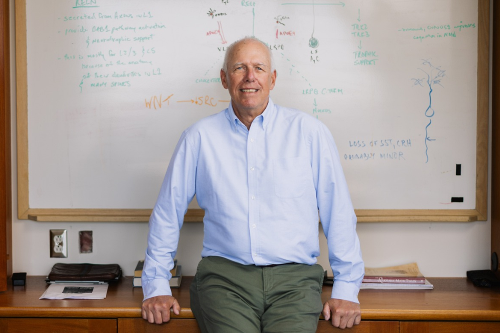
Nathaniel Heintz has spent decades building tools to peer inside individual brain cell types — and is now using those tools to understand some of the earliest changes that occur with Huntington’s and Alzheimer’s diseases. (Credit: Roshni Khatri)
An adult human brain contains roughly a hundred billion neurons—more than the number of stars in the Milky Way galaxy. Among these neurons is a vast diversity of cell types, with some cells carrying out completely different roles in the brain than other cells.
This immense complexity is what makes studying brain diseases, like Alzheimer’s and Huntington’s, so challenging. Scientists must figure out not only what goes wrong to trigger disease, but also in which cell types—and how that affects nearby cells.
For most of the history of neuroscience, researchers looked at a mishmash of isolated brain cells in big bulk experiments. That meant they couldn’t always detect what was happening in one particular type versus another, or understand how the spatial organization of cells played a role in brain function and disease development. Nathaniel Heintz, professor and head of the Laboratory of Molecular Biology, is working to change that.
Heintz has spent several decades building new tools to isolate specific cell types from human brain tissue, profile their molecular activity, and compare what’s happening in healthy brains versus those with disease. That work led to an entirely new kind of Parkinson’s disease drug, which just succeeded in phase 3 clinical trial. But Heintz says that’s just the beginning. His lab is now discovering that this data on individual cells can reveal some of the earliest changes that occur with Huntington’s and Alzheimer’s diseases—changes that happen only in specific cells and specific locations.
We spoke to Heintz, who also directs the Fisher Center for Alzheimer’s Disease at Rockefeller, about what his methods have revealed and what it will take to turn those results into treatments.
How much do brain cells differ from each other?
Enormously. The brain is one organ, but the cells in it look different from each other, fire differently, perform different functions, and certainly contribute differently to disease. My lab’s earliest experiments told us that there were one or two hundred genes that completely vary in activity between cell types; now we think there are closer to five hundred genes that really give each brain cell its particular properties.
And certain cell types are more vulnerable than others. So those genes that vary between cell types are precisely the ones you want to study if you’re trying to figure out why some cells get sick and others don’t. If you just look at bulk tissue, you won’t figure out those trends that are cell-type-specific.
For decades, scientists could only study brain cells as they were lumped together in tissue. Why was it so difficult to isolate different types of cells?
Even a small piece of brain tissue—a cube that’s one millimeter on each side—contains about ten million cells from more than a dozen cardinal cell types. If you want to understand what’s happening in one of those cell types, you have to be able to pull just those cells out of that ten-million-cell mixture.
We developed a method called fluorescence-activated nuclei sorting, or FANSseq (or NETSseq in industry settings). Every cell type, in its nucleus, has different proteins being used to regulate gene expression—that’s why it’s a different type. So we make fluorescent probes that latch on to proteins or RNAs that are unique to every type. Each set of probe pulls the entire set of nuclei for each cell type it targets from this complex mixture.
Once you have those nuclei, you can measure which genes are being transcribed into RNA, and how the structure of the DNA varies around different genes. You can cross-correlate these different levels of data to figure out whether a particular gene is really important in that cell type.
How do you turn all that raw data into disease insights?
Degenerative brain diseases unfold over decades. By the time someone dies from one of these diseases, the brain is so affected that you can’t really infer what happened at the start of the process. Tracing back those very first changes could help us pin down how one might prevent or stall the development of the disease.
We’ve been lucky enough to get postmortem brain samples from brain banks all over the world. These come from generous people and their families who dedicated their organs to science prior to death from Alzheimer’s, Huntington’s, Parkinson’s, and other causes. Using FANSseq, we can pull out specific cell types from those samples and compare them, gene by gene, to the same cell types from healthy brains. This lets us see which cell types are actually impacted.
We’ve discovered that in Huntington’s disease, for instance, there are distinct ways that gene regulation changes very early in disease—again, only in a few cell types. My lab uncovered why that gene change is detrimental in certain cells, while other cell types are spared. That has pointed us toward some possible ways to reverse the disease process specifically in those affected cell types.
You’ve recently started looking at Alzheimer’s disease. What have you found there?
We’re still very early in applying our tools to Alzheimer’s but I think our work is already very revealing. The classical hallmarks of Alzheimer’s—the amyloid plaques and tau tangles that you see in a diseased brain—are what most people focus on. But we think those events are actually quite late in the molecular cascade. By the time you have significant amyloid deposition, a great deal has already gone wrong upstream. The cells that were going to be most vulnerable have already been struggling for years.
What we want to find are those earlier events—any changes happening in specific cell types before the plaques and tangles appear in them.
If we can identify which circuits are impacted first, and which steps in those circuits break down earliest, that opens up two different therapeutic strategies. One is to intervene in the molecular cascade itself, trying to prevent or slow those early changes. The other is a kind of circuit compensation; if there’s a circuit where A leads to B, C, D, E, and F, and you discover that step E in a circuit is failing, you might be able to find a way to directly stimulate step F and restore function without needing to fix the underlying problem. That’s actually how some of the most successful neurological therapies for conditions like Parkinson’s or epilepsy work.
I think both of these strategies are important to pursue for late-onset diseases like Alzheimer’s and Huntington’s because even slowing progression significantly would be transformative for patients and families.
What does it take to move basic science observations toward new therapies for people with disease?
It requires a great deal of humility. You can have a very compelling hypothesis, supported by very good data, and still be wrong about whether intervening at a particular point in the disease cascade will help patients. The only way to know for certain is to test it in humans, and getting to that point is an enormous undertaking.
One of the challenges specific to brain disease is that you can’t model the full process of a degenerative disease in a mouse. Mice don’t live long enough, and their brains are different enough from ours. But what you can do is test specific hypotheses; if we think a particular epigenetic change is driving disease progression in medium spiny neurons, we can engineer a mouse with that change in those cells and see what happens. That kind of approach gives you one piece of the puzzle at a time.
So what you end up with is a cascade of hypotheses, each tested individually in mice or cells, that you’re stringing together into a compelling strategy that is worth the investment it takes to move it toward the clinic.
I’ve been through this process already. Using some of our earlier tools we figured out which circuits in the brain are impacted by Parkinson’s disease and how to compensate for that lost function. Now, the drug based on that work, called solengepras, has succeeded in a phase 3 trial, giving people with Parkinson’s more time each day with their symptoms under control and fewer side effects than existing drugs. At the same time, the study showed significant quality of life improvements, for instance with cognition and mitigating the sleepiness that comes not only from the disease itself but also from drugs currently used to treat it. While some of these positive effects were unexpected, overall, that’s exactly what you’d expect to see with a more targeted drug that comes out of studying very precise brain circuits. But it took decades to get from our first observations to the drug.
When that process is so long and unpredictable, what’s kept you excited about this work for so many years?
It’s the joy of teamwork and discovery as a team, really. If I didn’t have such wonderful students and postdocs and staff, and this collaborative nature across Rockefeller, I think it would have been hard to remain persistent in the face of such an incredibly difficult scientific challenge for so long. For me, these complex problems are great to tackle because you get to bring together teams of scientists who are exploring many different angles to get to a common answer. Over the past couple of decades, the cross fertilization of ideas with my Rockefeller colleagues Ines Ibanez-Tallon, Jeff Friedman, Titia de Lange, Bob Darnell, Mary Beth Hatten and the late Paul Greengard, when my thoughts were just the shadow of a concept, has been critical for expanding my horizons both technically and scientifically. I’m at the point in my career where it’s also about being able to take everything we’ve discovered and use that to explore new ways to help patients. Basic research is essential, in part because you cannot get to new therapies without it. And it should be noted that foundation support has been critical all throughout my research. Howard Hughes Medical Institute, the CHDI Foundation, The Fisher Center for Alzheimer’s Disease Research and Rockefeller have all been tremendously important for both enabling the research and providing the environment and colleagues for me to succeed.
We now know that we’ve got an approach that produces clinically relevant results. From here on out, I’m excited to focus on translating what we’re learning into the possibility of generating new treatments for these and other neurological disorders.