Novel tool gives researchers a better view of brain wiring
 COPY LINK
Key takeaways
- Electrical activity of neurons in the brain unfolds across vast interconnected networks so quickly that existing imaging tools have struggled to capture its underlying signals.
- A new imaging platform, FlatMux, overcomes this challenge by imaging electrical activity from about 200 neurons at once, deep in the mammalian brain and across multiple layers.
- This tool could ultimately allow scientists to visually trace how signals move through circuits, study the coordinated activity underlying various behaviors and cognitive function, and distinguish different cell types by their electrical signatures.
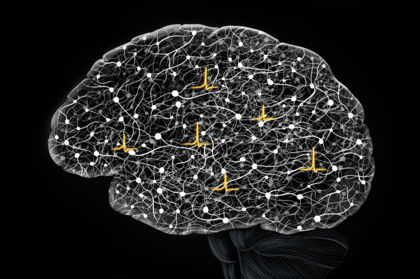
FlatMux captures electrical activity optically from nearly 200 genetically defined neurons across new depths in brain circuits, revealing how signals flow, cells connect, and computations emerge in a living mouse brain. (Image courtesy of the Vaziri lab)
The brain is a vast, densely interconnected network of neurons computing in parallel. Watching these computations unfold in real time could reveal how the brain processes sensation and guides behavior. But the electrical signals governing the network are fast, faint, and notoriously hard to capture in living tissue.
Now, a new study published in Nature Methods describes a two-photon imaging platform that captures electrical activity from almost 200 neurons at once, across cortical layers and at unprecedented depths and speeds, opening a window onto how information moves through living brain circuits. By minimizing the amount of energy needed to reliably detect the activity of a single neuron and devising a scalable strategy to optically scan laser pulses across the sample at a rate of 150 million samples per second, the platform, dubbed FlatMux, sets new benchmarks in the field, enabling recordings from nearly 200 neurons within one or several planes at once, at depths up to 500 micrometers, and at frame rates of up to 2,000 per second. With both the sensitivity and the speed that this tool offers, researchers can now begin mapping neurons that are functionally connected to and causally interact with one another, enabling a new understanding of how the brain’s wiring and function are giving rise to the brain’s cognitive capabilities. Moreover, unlike methods using electrode probes, the direct imaging of electrical activity via fluorescence allows for simultaneous inference of the location and the cell types of recorded neurons.
“For a long time the field has been focused on understanding the brain in terms of response properties of individual neurons. Our imaging platform makes it possible to instead investigate how various brain functions could be the result of information processing by a highly interconnected system where computation is distributed across a large network of neurons,” says Alipasha Vaziri, head of the Laboratory of Neurotechnology and Biophysics at Rockefeller.
Densely interconnected
Existing imaging tools have been hampered by different limitations. As far as optical tools are concerned, when trying to capture the densely interconnected system of brain cells, scientists long relied on calcium imaging, which uses fluorescent sensors to track calcium surges associated with neurons firing. But calcium signals are only an indirect measurement of electrical activity. In addition, they are much slower than the millisecond electrical impulses that brain cells use to communicate, so these techniques would miss key features of neural computation, including the signals that shape activity before a neuron fires (so-called sub-threshold responses).
In more recent years, researchers turned instead to genetically encoded voltage indicators, or GEVIs—fluorescent sensors embedded in cell membranes that report voltage directly. But GEVIs have their own challenges. Their signals are faint, about 100 times faster than calcium signals, and notoriously difficult to capture deep in living brain tissue. Imaging them at depth requires two-photon microscopy, a laser-based technique that can peer into scattering tissue but brings its own hard physical limits: too little laser power produces unusable signals, while too much can heat tissue or destroy sensors. Traditional two-photon systems are also highly inefficient in how they deliver light, limiting how many neurons can be recorded and for how long.
Vaziri set out to overcome such limitations, devising a tool that better captures the complex nature of brain activity. “We needed to come up with a system that makes excitation as efficient as it can be,” Vaziri says. “By maximizing the fluorescent photons we get out for the excitation photons we put in, we are able to look at as many neurons as possible.”
Rethinking light
To accomplish this, Vaziri and colleagues would need to rethink light delivery itself. They built a custom two-photon imaging platform, called FlatMux, that utilizes an optical cavity. The new platform splits a laser beam into many precisely timed pulses, which are delivered to a series of distinct, non-overlapping points in the tissue in a highly coordinated way. This is in contrast to the traditional approaches that rely on mechanical sweeping that results in laser pulses oversampling each region during a scan. Their approach not only allows for faster scanning but also makes the system far more efficient, generating stronger signals from less light, while also reducing tissue heating and undesirable artifacts. With FlatMux, researchers can image larger groups of neurons, deeper in the brain, and fast enough to capture the millisecond electrical events neurons use to communicate. Moreover, the design of the system is scalable which means that, as GEVIs continue to improve, FlatMux will be able to reallocate realized gains in energetic resources to record from even larger neuronal populations while maintaining the same speed and without further increases in power.
The team then paired FlatMux with custom computational tools designed to distinguish true neural activity from background noise. Together, the hardware and software work as a single platform: one maximizes how much information can be extracted from every photon sent into the brain, while the other reconstructs the fleeting electrical signals hidden in that data. Then, to test it in realistic conditions, the team used the system in awake mice running on a treadmill that were exposed to whisker stimulation, showing that the system could capture faint voltage signals during such stimulation as well as during spontaneous behavior.
The result was a system built not just to image more neurons, but to capture neural computation in action. And the payoff was dramatic. FlatMux recorded almost 200 neurons simultaneously, reached depths of 500 micrometers, captured activity at up to 2,000 frames per second, and even imaged two cortical layers at once, allowing researchers to watch signals move through the layered cortical circuitry in real time. In a high-sensitivity mode, the system also detected signals too weak to trigger firing but critical for revealing the neurons that are synaptically coupled to one another, raising the possibility of mapping functional connections optically and in a high-throughput manner rather than one cell pair at a time.
With FlatMux, it may finally be possible to move beyond snapshots of isolated neurons to capturing computations as they unfold across distributed networks in real time. That could help researchers trace how signals move through circuits, study the coordinated activity underlying various behaviors such as decision-making, and potentially even transform the burgeoning field of connectomics. Its millisecond precision may also help researchers distinguish different kinds of brain cells by their electrical signatures, opening a new way to classify cell types by function.
“A lot of effort has been put into the goal of understanding the brain-wiring diagrams of model organisms,” Vaziri says. “The FlatMux platform could be used to infer how neurons are synaptically connected.”
More broadly, FlatMux points toward a shift in neuroscience, from sampling fragments of activity to capturing how living brain circuits compute in real time. “The brain is not only a biological organ but also an information-processing system, and I’ve always been excited about figuring out its underlying computational principles,” Vaziri says.
“When we build these kinds of tools, it’s not just about overcoming an engineering challenge. It’s about enabling neuroscience to ask deeper questions about how the brain computes.”

Key takeaways
- Electrical activity of neurons in the brain unfolds across vast interconnected networks so quickly that existing imaging tools have struggled to capture its underlying signals.
- A new imaging platform, FlatMux, overcomes this challenge by imaging electrical activity from about 200 neurons at once, deep in the mammalian brain and across multiple layers.
- This tool could ultimately allow scientists to visually trace how signals move through circuits, study the coordinated activity underlying various behaviors and cognitive function, and distinguish different cell types by their electrical signatures.