Getting an unprecedented view of the molecular machines of life
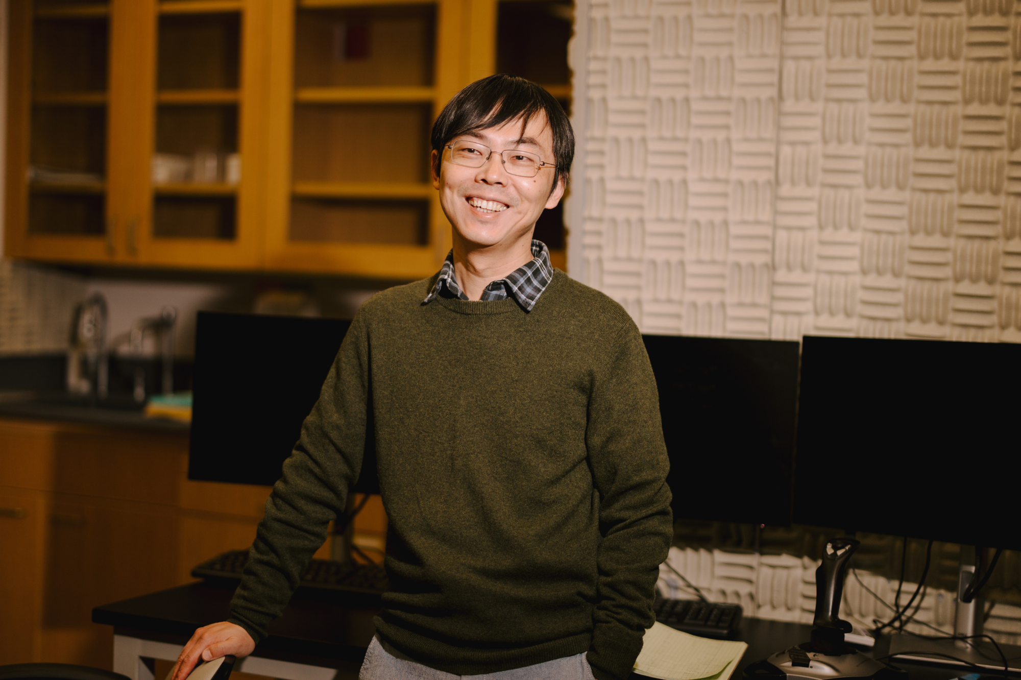
Shixin Liu is pioneering new ways of studying the tiny proteins that copy and read DNA in living cells. (Credit: Roshni Khatri)
Inside every cell of your body, tiny molecular machines are whizzing along strands of DNA that contain the entire blueprint for making and operating a human body. These nanoscale proteins copy DNA and transcribe genes into the shorter bits of RNA that can encode new proteins to keep cells functioning. Though minuscule, these machines conduct their functions with remarkable accuracy and efficiency—generating considerable forces to tug and twist DNA or clear roadblocks therein—all in the midst of a chaotic cellular environment. They rarely make missteps, but when they do, the health consequences can be dire.
Shixin Liu, associate professor and head of the Laboratory of Nanoscale Biophysics and Biochemistry at Rockefeller University, wants to understand how exactly these machines work. To do that, his lab develops technology to observe individual molecules in action, capturing a record of each machine’s movements. They have also devised inventive ways to apply physiologically relevant amounts of force to these proteins and test how the molecules respond.
We spoke to Liu about what makes these machines so fascinating and why studying them could lead to a new understanding of diseases from tuberculosis to cancer.
What are the molecular machines that your lab studies?
The two main processes we focus on are DNA replication (how a cell copies its genome before it divides) and transcription (how DNA is copied to RNA that can be used to make proteins). The machines that carry out these processes are staggeringly complex, with many moving parts that all must be precisely coordinated. They are also distinctly mechanical—which means they’re converting chemical energy into physical forces as they work.
What makes them especially fascinating is the environment they work in. At the nanometer scale, where these machines operate, random thermal fluctuations are constantly knocking everything around. Yet they make remarkably few errors. That’s phenomenal; it’s also hugely consequential, because when these machines malfunction, cancer, developmental disorders, and neurological diseases can arise.
How do you study the intricate movements of these tiny proteins?
There are two components of our approach: fluorescence detection and force manipulation. With fluorescence detection, we label a molecule with a fluorescent tag that lets us visualize its movements and conformational changes in real time. With force manipulation, we use focused lasers called optical tweezers to measure forces the molecules generate and energies they consume.
My lab combines these tools, which is powerful because you can simultaneously perturb the system with force and visualize its behavior. I like the analogy that if you only use fluorescence, you’re watching with your hands tied behind your back. If you only use force manipulation, you can pull on things, but you’re blindfolded. If you can do both—see and manipulate at the same time—you understand a system much better.
What are you trying to understand about these machines?
The overarching question is how they orchestrate the assembly and activity of each component to ensure optimal function. This question cannot be answered by traditional genetic and biochemical approaches because of the dynamic and stochastic nature of molecular events, but our single-molecule methodology is ideally suited for tackling it. Also, these machines do not operate in isolation—they are simultaneously working in close proximity on a crowded genome and constantly bumping into each other. How do cells manage this traffic and make sure every machine is doing its job or even harness the traffic to its advantage?
The other question we can answer exceptionally well is how fast these machines move on DNA. Speed matters greatly—take the transcription machines as an example. When they move too quickly or too slowly, not only does the process of RNA synthesis itself go awry, other processes coupled to transcription—like how RNA molecules are processed and how DNA is packaged—become dysregulated as well, which can contribute to human disease.
What have been some of your most surprising discoveries?
The common theme across what we’re finding is that these proteins are more versatile than we thought. Many do more than one job. We often think of the cell as a highly evolved factory where each machine has its specific task, but biological molecules are far more adaptable—they carry out different functions, sometimes makeshift ones, which makes the apparatuses more robust.
One example involves a ring-shaped protein called a sliding clamp, which encircles DNA to help the polymerase enzyme copy it. Another protein, the clamp loader, opens the ring and snaps it onto the DNA. For a long time, people thought that after loading the clamp, the loader dissociates and its job is done. We recently found—through direct single-molecule visualization—that the loader actually stays around, fulfilling a separate role which is to keep the sliding clamp tightly associated with the polymerase during DNA synthesis. When the clamp loader isn’t there, genome replication cannot be properly carried out especially when the cell is under stress.
How do your experiments shed light on disease or point toward new drug candidates?
The core components of the DNA replication and transcription machinery are so fundamental that mutations in them are usually lethal—cells can’t survive without them. But many of the regulators and accessory proteins are frequently mutated in disease. When these molecular machines fail to coordinate internally or with each other, you get aberrant gene expression or DNA damage. That means that many of the molecules we study are actually good drug targets for treating disease such as cancer and neurological disorders.
For instance, we’ve studied one protein called MeCP2 which helps control which genes get turned on and off in brain cells. Mutations in MeCP2 are known to cause Rett Syndrome, a devastating neurodevelopmental disease that primarily affects young girls. We discovered previously unknown ways that MeCP2 interacts with DNA, which could open up new opportunities to treat Rett Syndrome, which still lacks a cure.
In another project, we studied transcription in Mycobacterium tuberculosis and discovered that the machinery pauses frequently, in a mode of regulation not seen in many other bacteria. Such pausing could be a vulnerability that can be exploited for antibiotic development.
Where is this research headed next?
In eukaryotic cells like ours, DNA isn’t just a bare strand—it’s wrapped tightly around proteins in structures called nucleosomes, which package the majority of the genetic material. My lab is moving toward studying how machines process this packaged DNA rather than just bare DNA. We want to see how they navigate through nucleosomes—removing these roadblocks but then reassembling them again once they’re past. It’s a daunting but critical task for the molecular machines, given the rich epigenetic information stored in the nucleosomes. We’ve built up the groundwork to visualize these processes for the first time.
New automation and machine learning tools that we are developing will allow us to perform experiments at high throughput and extract more insights from the information-rich datasets that we collect. We are also striving to continue increasing the complexity of our experimental systems so the measurements can better mimic what is happening in the cellular environment. After decades of technological advancement made by the field, we’ve reached the point where these powerful tools can be used to uncover fundamental biological processes that have never been seen before. The potential for where we can go from here is enormous.