The challenge for cell biologists in the mid-twentieth century was not unlike the challenge for astronomers. Belgian biologist Albert Claude put it, “the cell was as distant from us as the stars and galaxies.” At that time, the inner workings of the cell, millions of times smaller than the head of a pin, were invisible to even the best light microscopes. The electron microscope, first invented by German engineers in 1931, proposed a solution to the problem of cell biology with its much higher magnification power. But preparing live cells for the technology proved an obstinate hurdle. In the 1940s and 1950s, work by Belgian biochemist Christian de Duve helped modernize the field of cell biology by removing this hurdle. For this work and his subsequent discoveries of the structure and function of cellular components, Dr. de Duve shared the 1974 Nobel Prize in Physiology or Medicine.
To microscopists in the early 20th century, the sheer number of components in a living cell made it appear a jumbled, indecipherable mass. In 1930, Dr. Claude, working at The Rockefeller Institute for Medical Research, devised a technique, later perfected by Dr. de Duve and others, to separate the functional parts of a cell and thus allow for clearer examination with powerful electron microscopes. Called cell fractionation, Dr. Claude’s method involves grinding cells in order to break the cell membrane and then sorting the released contents by mass and weight through precisely calibrated cycles of centrifugation.
Using Dr. Claude’s cell fractionation technique, Dr. de Duve discovered lysosomes, which act as the cell’s clean-up crew, in 1949 while working at the Catholic University of Louvain in Belgium. Dr. de Duve and his colleagues were actually trying to measure certain enzymes in the centrifuged cell fractions in order to determine their intracellular localization. They noted that one of the enzymes — acid phosphatase, which had been included largely for control purposes — was only one-tenth as active in this experiment as it had been in previous demonstrations using the more drastic homogenizing action of the Waring Blender. In further experiments, Dr. de Duve found that in living cells this enzyme is largely or entirely confined within small bag-like particles, which were later found to contain more than a dozen different hydrolytic enzymes capable of acting on all major constituents of living matter. Considered as a group, these enzymes could have only a digestive function, and hence, the particles containing them were called lysosomes (lytic bodies). In 1955 Dr. de Duve and Alex B. Novikoff of the Albert Einstein College of Medicine identified lysosomes visually when they obtained the first electron micrographs of cell fractions containing partially purified lysosomes.
As Dr. de Duve’s work shows, cell fractionation provided an unprecedented opportunity to peer inside the once-cloistered world of the cell, and the main players in the drama — Dr. de Duve, Dr. Claude at Rockefeller and Dr. Claude’s graduate student George E. Palade — were prolific in their examination and biochemical analysis of each distinct constituent of various cell types. Their work over the next few decades resulted in the first functional map of the cell, and what they revealed was an entire microcosm: In addition to lysosomes, they discovered mitochondria, power plants of the cell; vacuoles, which police harmful elements; ribosomes, which produce worker-proteins; and many other, even smaller subcellular components.
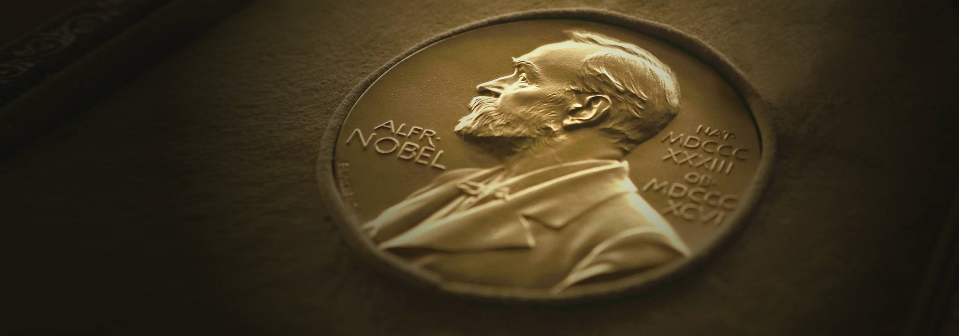
By the early 1950s, cell biology was a recognized field. In 1955, The Journal of Cell Biology was established at The Rockefeller Institute, and in 1960, the American Society for Cell Biology was founded. Drs. de Duve, Claude and Palade, who would together share the 1974 Nobel Prize, were directly involved in these and other ventures of the burgeoning new field they had largely created. Dr. Claude, in his Nobel acceptance speech, described the three men’s discoveries together: “We have entered the cell, the mansion of our birth, and started the inventory of our acquired wealth.”
CAREER
Born outside London in 1917, Dr. de Duve was raised in Antwerp, Belgium. Following his medical degree, which he earned from the Catholic University of Leuven in 1941, the lack of available research positions brought on by World War II led him to seek further education in chemistry and biochemistry, for which he studied at Leuven Cancer Institute, the Medical Nobel Institute and Washington University in St. Louis. He returned to the Catholic University of Leuven in 1947, becoming professor in 1951. He joined The Rockefeller Institute as professor in 1962 and from then split his time between his laboratories in New York and Leuven. In 1974, Dr. de Duve founded the International Institute of Cellular and Molecular Pathology, now known as the de Duve Institute, in Brussels, and he served for several years as its president. He became emeritus professor at Leuven in 1985 and at Rockefeller in 1988. Dr. de Duve authored three books: A Guided Tour of the Living Cell, Blueprint for a Cell, and Vital Dust.