Hilda Amalia Pasolli, Ph.D.
Research Associate Professor
Director
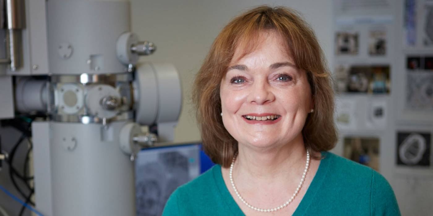
Pasolli re-joined The Rockefeller University in 2019 as the director of the Electron Microscopy Resource Center, which provides expertise and training in electron microscopy, including sample preparation and image analysis, for a variety of specimens.
A cell biologist and electron microscopist by training, she uses a wide range of electron microscopy methods to study the ultrastructure of a variety of cells and organisms, ranging from bacteria to the complex structure of mammalian tissues. She interprets ultrastructure of cells and tissues and also devises novel and improved protocols for procedures.
After completing her undergraduate degree at Cordoba National University in her native Argentina, she joined the Center for Electron Microscopy (CEM) there for her doctoral studies characterizing pituitary cell types. She then pursued postdoctoral studies in neurobiology at the University of Heidelberg in Germany before returning to the CEM as assistant professor and investigator of the National Council for Scientific and Technical Research. She then worked for 12 years in Elaine Fuchs’ lab at Rockefeller, where she applied her electron microscopy skills to studying the ultrastructure of skin in a variety of mouse models. In 2014 she joined the Howard Hughes Medical Institute’s Janelia Research Campus in Virginia, where she gained experience in correlative light-electron microscopy methods and cryofixation of cells and tissues.
She has received multiple awards from the Microscopy Society of America for her application of electron microscopy to the biological sciences. Her work has been published in two textbooks, and laboratories all over the world request her advice on electron microscopy, especially for studies of skin ultrastructure.