Expert in cryo-electron microscopy to join Rockefeller faculty
Thomas Walz, a structural biologist who uses cutting-edge electron microscopy techniques to better understand processes involving biological membranes, will join Rockefeller’s faculty as a tenured professor on September 1. As head of the Laboratory of Molecular Electron Microscopy, Walz will take advantage of the university’s recently acquired cryo-electron microscopes, which can capture molecular structures in unprecedented detail, to advance his research on macromolecular complexes and proteins embedded in cellular membranes.

Thomas Walz
“Tom has unparalleled expertise using sensitive new techniques to explore the architecture of biologically important molecules, and as a result, their function,” says Marc Tessier-Lavigne, the university’s president. “His addition to our faculty will further strengthen our thriving team of structural biologists, and his deep knowledge of modern electron microscopy tools will make him an indispensable colleague for many within our community, helping us make the most of our recent investments in this technology.”
Walz began his scientific career studying the structures of proteins embedded in the membrane that surrounds cells, which is composed of two layers of molecules known as lipids. As a Ph.D. student at the University of Basel’s Biozentrum in Switzerland, he began work on the structure of aquaporin-1, a protein that forms a selective channel to allow water to travel in and out of cells. The structure resolved a long-standing riddle as to how aquaporins can efficiently conduct water while they are impermeable to protons, which should be able to move along with the water.
But long before that, the seed for his investigation of the unseen world was planted when, as a university student, he looked through an electron microscope at a drop of what appeared to be clean water but contained particles that looked like capsules designed for landing on the moon. These were viruses with their protein shells, called capsids.
“It was like looking into a different world. Some people are fascinated by the universe, I became fascinated by the very small structures that can’t be seen with the eye alone,” Walz says. “Because I am a very visual person electron microscopy made sense for me, because you always see what you are studying.”
Following his Ph.D. work, Walz moved to the University of Sheffield in the United Kingdom, where as a postdoc, he determined the two-dimensional structures of three photosynthetic complexes of membrane proteins from the bacterium Rhodobacter sphaeroides. After joining the faculty of Harvard Medical School in 1999, he continued studying the structures of other aquaporins, including aquaporin-0, a water channel in the eye’s lens that also acts as an adhesive for cell membranes, helping to form connections between cells.
“The images we obtain of aquaporin-0 had a high enough resolution that they also revealed the lipids in the surrounding membrane. That started to get me interested in how proteins interact with lipids and how proteins and lipids accommodate each other,” Walz says.
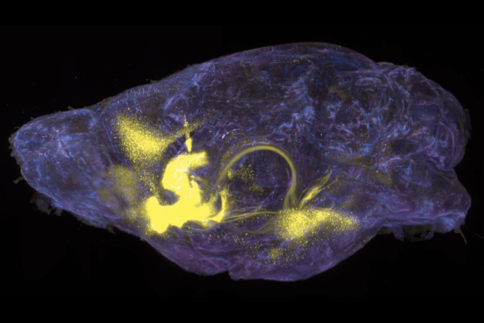
This interplay remains poorly understood. Proteins embedded in the cellular membrane are responsible for carrying out the membrane’s functions: relaying signals, allowing for cargo transport, catalyzing reactions, and mediating all interactions with the external environment and other cells. Studies continue to reveal the structures of these proteins, and as a result, how they carry out these activities. However, most of this structural work has been conducted on isolated membrane proteins in solution, without the lipid bilayer that is the membrane protein’s native environment. Meanwhile, cellular membranes contain thousands of different lipids, and it is being increasingly recognized that this diversity affects most membrane processes as well as the membrane proteins themselves.
Taking advantage of new tools in electron microscopy, Walz investigates the structure and function of membrane proteins within the context of the lipid environment and of macromolecular complexes, such as those that guide transport of cargo throughout the cell. In addition to ever more sophisticated software to calculate high-resolution structures from noisy images, one of the major developments he employs, direct electron detector device cameras, enables scientists to record images and movies with unprecedented contrast and to compensate for the inevitable movement of specimens that occurs at this scale. Walz also uses nanodiscs — small patches of lipid bilayer stabilized by a scaffolding protein — to study how lipids affect membrane protein structure and function. Because they recreate a membrane protein’s native environment, nanodiscs are a significant improvement upon the detergents traditionally used in electron microscopy.
Walz is already collaborating with a number of Rockefeller scientists including Roderick MacKinnon, Jue Chen, Seth Darst, Günter Blobel, Tarun Kapoor, and Sebastian Klinge. Walz will guide the structural biologists at Rockefeller to become independent in the use of cryo-electron microscopy, and he also looks forward to collaborating with biologists from other disciplines to address questions in their research fields that are best tackled by this approach.
“I am looking forward to joining Rockefeller’s thriving community of structural biologists. Both the state-of-the-art electron microscopy tools and the university’s small, collegial environment were crucial factors in my decision to move my lab,” Walz says.