New cryo-EM suite expands Rockefeller’s capabilities in structural biology
by ZACH VEILLEUX
Structural biology, in which scientists examine the shapes of specific proteins and protein complexes at a molecular scale, has driven some of biology’s most profound discoveries in the past decade, including insights into neurological signaling, pathogenic processes and DNA transcription. With the acquisition of sophisticated new cryo-electron microscopy tools, the university’s labs will be able to benefit from technology that allows for the visualization of three-dimensional structures of molecules and macromolecular complexes in solution.

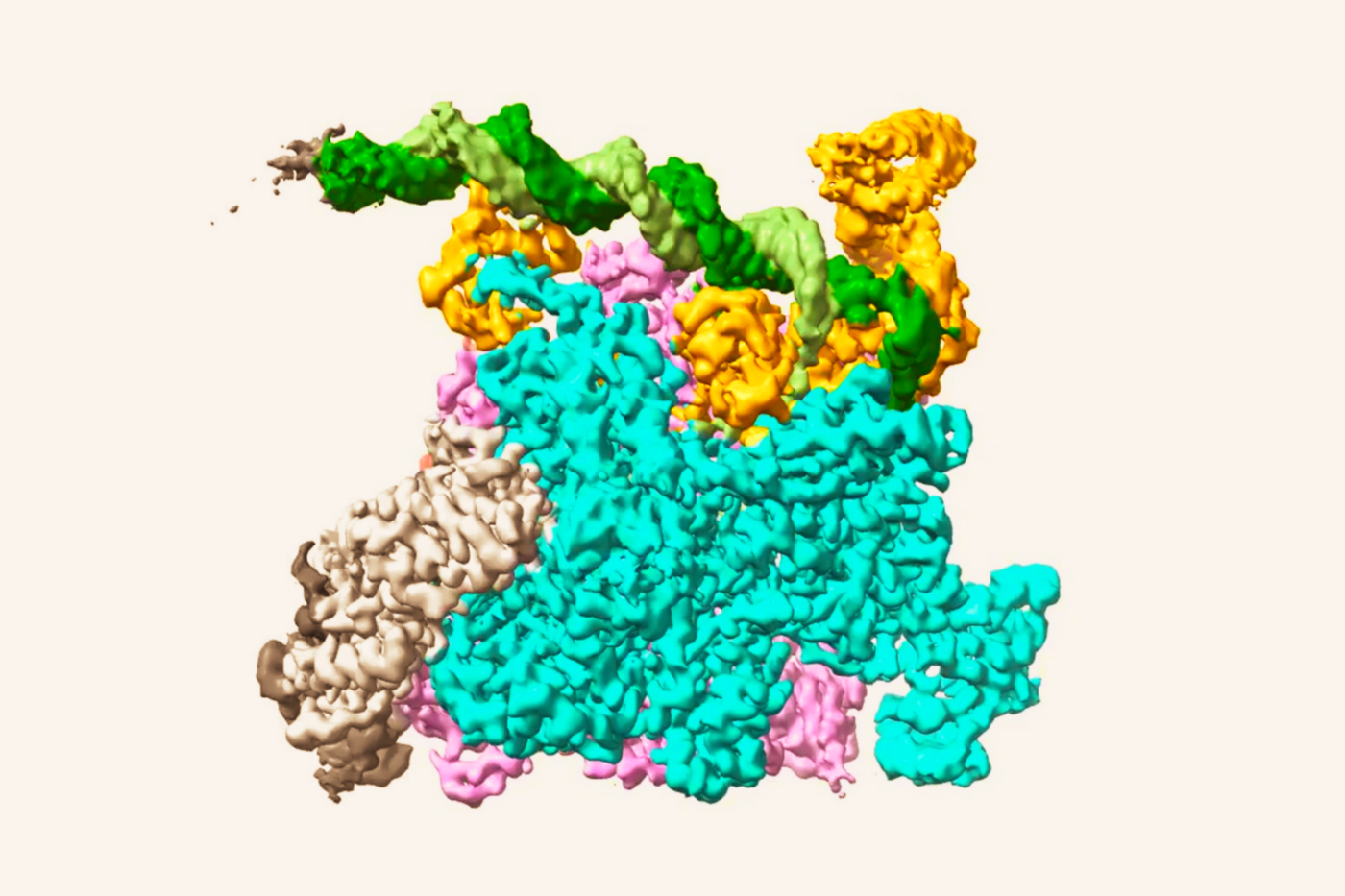
Science on Ice. A structure of an ion channel obtained using new cryo-electron microscopy technology by scientists at the University of California, San Francisco, where the technique was pioneered.
The new equipment, purchased in October with a grant from the EGL Charitable Foundation — thanks to Rockefeller Trustee Evelyn Gruss Lipper and her daughter Daniella Lipper Coules — will be installed on the C-level of Smith Hall in the coming weeks. It will be operated by a yet-to-be-hired microscopy specialist, and used by more than a dozen laboratories working in chemical and structural biology, molecular and cell biology, immunology, neuroscience and other fields.
A cluster of structural biologists — Roderick MacKinnon, Seth A. Darst, Jue Chen and Sebastian Klinge — has championed the initiative to purchase the equipment and is overseeing its implementation.
“The advances in the cryo-EM method are revolutionizing structural biology, allowing detailed structural analysis of proteins and other biological macromolecules, many of which can not be successfully visualized with existing technologies,” says Darst, who is Jack Fishman Professor and head of the Laboratory of Molecular Biophysics.
Although cryo-EM has been around for years, it usefulness in biology has been restricted by its inability to yield detailed molecular structures. Biologists interested in understanding the molecular structures of specific proteins or protein complexes have relied on other technologies, primarily x-ray crystallography. Not all biological molecules can be crystallized, however, and the crystallization process itself is both painstaking and unpredictable. Instead of crystals, cryo-EM can be used to examine uncrystallized biological samples at cold temperatures within thin sheets of ice.
A breakthrough in detector technology, made just last year, has dramatically improved the ability of cryo-EM equipment to capture fine structural information at scales as small as tenths of nanometers. The new detectors not only sense electrons with greater precision as they pass through cryogenically frozen samples, they also take advantage of computation methods to correct for disruptions in the data including the effects of motion caused by the electron beam itself.
By offering the possibility to determine the structures of protein complexes that have resisted other techniques, and to view variations in those complexes that may occur when they are active, cryo-EM has the potential to drive new discoveries throughout biology.
“Investing in cutting-edge technology such as cryo-EM is critical to maintaining Rockefeller’s competitive edge in the life sciences, and we are grateful to Evelyn Lipper and her family for providing the resources to make this acquisition possible,” says Marc Tessier-Lavigne, the university’s president. “Obtaining this technology now will assist a number of Rockefeller biologists — including non-specialists in chemistry and structural biology — who want to apply these techniques to their investigations of a wide range of scientific problems.”