Rockefeller's Cryo-EM Center enters a new chapter
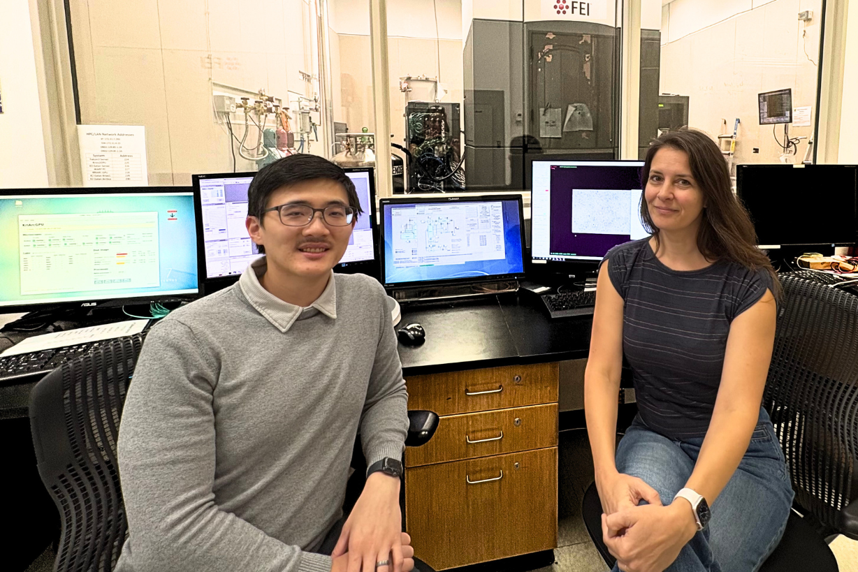
Honkit Ng and Johanna Sotiris
With expanded capabilities and new co-leadership, the Cryo-Electron Microscopy Resource Center continues to advance structural biology research at The Rockefeller University.
Since opening in 2016, Rockefeller’s Evelyn Gruss Lipper Cryo-Electron Microscopy Resource Center (CEMRC) has undergone a significant transformation. What began as a facility providing researchers with access to single particle analysis capabilities of the newly developed higher resolution cryo-electron microscopes has grown into a resource offering increasingly sophisticated technologies for investigating biological structures at near-atomic resolution.
The Center has now entered a new chapter, with Johanna Sotiris and Honkit Ng taking on co-leadership. Together, they oversee the Center’s operations and scientific support, combining complementary expertise to help Rockefeller investigators make the most of its technologies.
Over the last decade, the Center expanded significantly, adding a second Titan Krios microscope, upgrading all three electron microscopes, and introducing new capabilities in focused ion beam (FIB) milling and cryo-correlative light and electron microscopy (cryo-CLEM). These advances have broadened the range of biological questions researchers can address, including the study of molecular structures within their cellular environments.
For Sotiris, whose responsibilities include microscope operations, sample preparation, and facility management, ensuring that researchers can use these sophisticated instruments effectively is a central priority.
“Our goal is to make these technologies accessible to researchers, whether they are experienced cryo-EM users or just beginning to explore what the methods can offer,” says Sotiris. “We work closely with them to prepare samples, develop imaging strategies, and obtain the best possible data.”
Ng brings expertise in cryo-electron tomography, advanced imaging workflows, and the computational infrastructure required to manage the large datasets generated by modern microscopes. He also coordinates with Rockefeller’s High-Performance Computing team and the New York Structural Biology Center (NYSBC), helping connect investigators with additional resources and expertise.
“Cryo-EM is evolving rapidly, and so are the possibilities for researchers. By integrating new imaging approaches with efficient data collection and processing workflows, we can support increasingly complex projects and help investigators tackle questions that were previously difficult to address.”
A recent example of the Center’s contribution to Rockefeller science comes from the Laboratory of Molecular Biophysics, headed by Seth Darst. In a June 2026 study published in Proceedings of the National Academy of Sciences (Dhingra et al., 2026), researchers used cryo-EM to reveal how experimental antibiotic compounds interfere with the molecular movements of RNA polymerase, an enzyme essential for gene expression. The findings offer insights that could inform future antibiotic development. The study acknowledged CEMRC staff for their support with cryo-electron microscopy instrumentation and sample handling.
As the CEMRC approaches its tenth anniversary, its expanding technologies and collaborative approach continue to strengthen Rockefeller’s structural biology research, with Sotiris and Ng jointly guiding its next phase.
To learn more about the services offered by the Evelyn Gruss Lipper Cryo-Electron Microscopy Resource Center, visit rockefeller.edu/cemrc/
To read more news about the Scientific Resource Centers, visit the Resource Center News page.
Reference:
Dhingra, Y., Landick, R., Campbell, E. A., & Darst, S. A. (2026). RNA polymerase inhibitors reveal active-site motions essential for the nucleotide addition cycle. Proceedings of the National Academy of Sciences, 123(27), Article e2609228123. https://doi.org/10.1073/pnas.2609228123