Stem cell model offers first glimpse of early human development
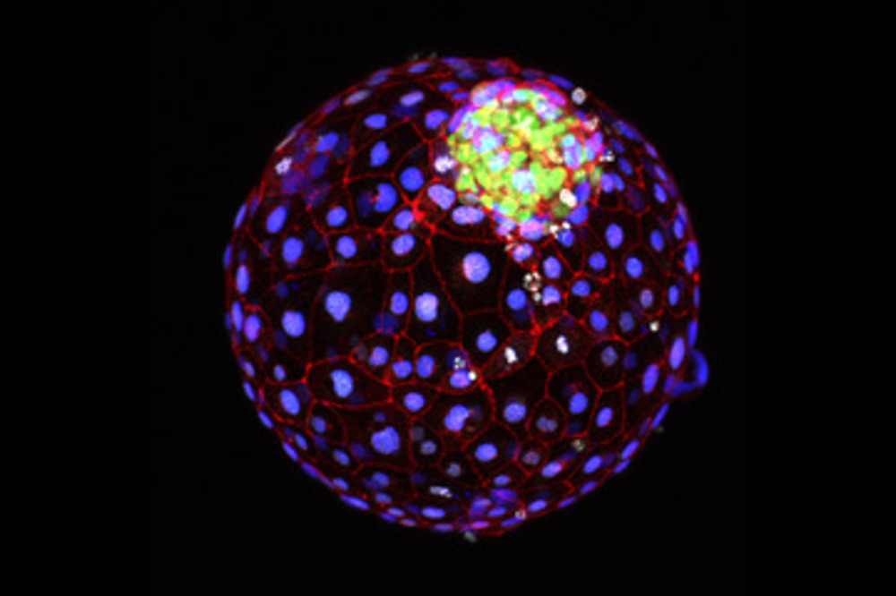
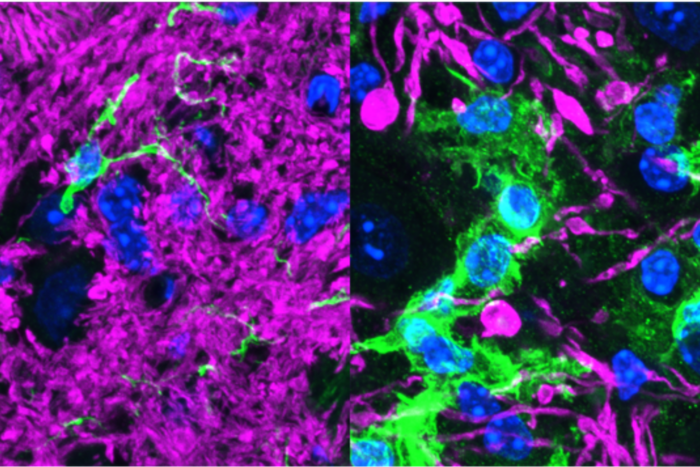
A blastoid, a stem cell model system that allows scientists to study the nuances of human gastrulation. Credit: Brivanlou Lab
It’s one of life’s most defining moments—that crucial step in embryonic development, when an indistinct ball of cells rearranges itself into the orderly three-layered structure that sets the stage for all to come. Known as gastrulation, this crucial process unfolds in the third week of human development. “Gastrulation is the origin of our own individualization, the emergence of our axis,” says Rockefeller’s Ali Brivanlou. “It is the first moment that separates our heads from our behinds.”
Observing the molecular underpinnings of this pivotal event would go a long way toward helping scientists prevent miscarriages and developmental disorders. But studying human gastrulation has proven both technologically difficult and ethically complicated, and thus current approaches have had limited success in expanding our understanding of early human development. Now Brivanlou and colleagues have demonstrated how a stem cell model system known as a blastoid can allow the study of the nuances of human gastrulation in the presence of pre-implantation extra-embryonic cell types. Their study, published in Stem Cell Reports, describes the scientific and clinical potential of this new platform.
“Gastrulation was a tremendous black box. We had never seen ourselves at that stage,” Brivanlou says. “This moves us closer to understanding how we begin.”
A better blastocyst
Prior to implantation, an embryo is a ball of about 250 cells organized as a blastocyst. This elusive ball of cells was difficult to study directly, so scientists developed blastoids—stem-cell-based blastocyst models. Blastoids can be cloned, experimentally manipulated, and programmed, allowing scientists to study identical blastoids over and over again.
The question was whether blastoids could gastrulate in vitro. Unlike a blastocyst in vivo, which rolls around in the uterus until it attaches to maternal tissue, blastoids were good at modeling the ball of cells from which life emerges, but it remained unclear whether this in vitro model could model later stages of human development. That is, until Brivanlou developed a platform to allow blastoids to attach in vitro, and thereby progress toward gastrulation.
“We were then able to see epiblast symmetry breaking, marked by BRA expression, for the first time with the high molecular resolution,” says Riccardo De Santis, a research associate in the Brivanlou lab and lead author on the study. “This allowed us to start asking more detailed questions about the earliest moments of life.”
With this unprecedented clarity, the team directly observed two key moments in gastrulation: the first epiblast symmetry-breaking event and the emergence of the molecular markers of the primitive streak and mesoderm upon in vitro attachment.
The primitive streak is a structure that marks the beginning of gastrulation and lays the foundation for the three primary layers of the embryo. One of those layers, the mesoderm, forms during gastrulation and gives rise to muscles, bones, and the circulatory system. The team discovered that, as early as seven days after attachment, they were already able to use molecular markers to detect the earliest signature of a nascent primitive streak and mesodermal cells.
To confirm their findings, the team also compared the blastoid results with data from in vitro attached human embryos and demonstrated that blastoids express the same genes in vitro that a regular embryo would at that stage in vivo, a strong demonstration of the power of blastoids as models for human embryonic development. Further highlighting the power of the lab’s in vitro attached blastoid system, the team then used it to demonstrate that pathways that regulate the rise of the primitive streak and mesoderm in vivo also regulate blastoids symmetry breaking in vitro—all with nothing but stem-cell-derived blastoid models.
Along the way, the team also demonstrated that gastrulation in vitro can begin at day 12, earlier than once thought. “This will change textbooks,” Brivanlou says. “We’ve contributed to redefining the molecular signature and timing of the onset of gastrulation upon in vitro attachment”.
Therapeutic possibilities
The results demonstrate that blastoids, when combined with the Brivanlou lab’s unique attachment platform, are now capable of conveying insights into early human development that have long been inaccessible. De Santis envisions a future in which blastoid-based research leads to advancements in diagnosing and treating developmental disorders, or offers insights into potential causes of early miscarriages during gastrulation.
“Many couples can’t have babies because the embryo doesn’t attach properly, and many miscarriages occur in the first few weeks of pregnancy,” De Santis explains. “We now have a model system that can help us understand the molecular mechanism that defines whether a pregnancy will be successful or not.” In the near future, De Santis hopes to combine this method with machine learning to help predict pregnancy outcomes and the trajectories of developmental disorders by observing how model blastoids built with particular genetic makeups fare in vitro.
“A better understanding of gastrulation—and the ability to study it with a reliable model system—impacts everything from survival of the fetus to autism to neurodegeneration.”