Rockefeller welcomes three new lab heads
by WYNNE PARRY
In the next five months, three new laboratories will open on campus, their research centering on cellular metabolism, biological membranes, and molecular motors. Two of the new faculty recruits are tenure-track candidates who emerged as finalists in last year’s open search. The third, Thomas Walz, joins Rockefeller as a mid-career hire.
“This has turned out to be an excellent year for faculty recruitment,” says Marc Tessier-Lavigne, the university’s president. “We set a very high bar for our new faculty, and we devote substantial resources to vetting the best candidates. It is immensely rewarding when those efforts pay off and we are able to welcome phenomenal scientists into our community.”
Kivanç Birsoy
Kivanç Birsoy studies the changes in cellular metabolism that occur in disease, including cancer. Currently a postdoc at MIT’s Whitehead Institute for Biomedical Research, Dr. Birsoy will relocate to Rockefeller in January and establish the Laboratory of Metabolic Regulation and Genetics.
“The complex metabolic pathways by which cells process nutrients have been well mapped at this point. However, little is known about how cells regulate their metabolism so as to adapt to environmental and genetic changes,” Dr. Birsoy says. “By exploring this form of regulation, ultimately, I hope to develop therapies for diseases involving alterations to metabolism.”
A native of Turkey and a Rockefeller alumnus, Dr. Birsoy was a Ph.D. student in Jeffrey Friedman’s lab, where he made a number of fundamental discoveries about fat cells. As a postdoc, his interest shifted to cancer. In order to study the metabolism of tumor cells, he designed and used new tools, including an instrument for mimicking the nutrient-deprived environment within some tumors.
Upon returning to Rockefeller, he will continue working on cancer, as well as two other categories of disease: mitochondrial disorders and metabolic disorders arising from genetic errors. He plans to use a combination of metabolomics and genetic tools, such as CRISPR-Cas9 genome editing technology, to examine how cells regulate their metabolism in disease conditions.
Because tumors are often cut off from an organism’s blood supply, cancer cells frequently lack the nutrients available to normal cells, and must alter their metabolism in order to proliferate. In some cases, they become dependent on certain nutrients that other cells are capable of manufacturing. As a postdoc, Dr. Birsoy’s work on cancer cells’ sensitivity to glucose led to the finding that certain genetic mutations can render cancer cells vulnerable to drugs known as biguanides, which are used to treat diabetes. He plans to build on this work by mapping out cancer cell dependencies on other nutrients, such as amino acids and lipids, while simultaneously looking for opportunities to exploit them for cancer therapy.
Dr. Birsoy’s second area of focus is on mitochondrial disorders, which occur when these energy-generating organelles within cells don’t work properly. This situation affects multiple systems within
the body, producing neurological and muscular problems. Current treatments are largely limited to managing the symptoms, rather than addressing the cause of the disease. Dr. Birsoy plans to investigate how specific mitochondrial dysfunctions affect cellular metabolism, and so produce various symptoms.
The third area of focus for the Birsoy lab will be rare genetic disorders that result in metabolic errors, in which metabolites accumulate to toxic levels. In organic acidurias, for example, patients are unable to break down amino acids, which build up in blood and are excreted in urine. These conditions can produce symptoms such as neurological damage, developmental delay, lethargy, and vomiting, and can lead to death. Dr. Birsoy aims to better understand the mechanisms by which metabolites such as amino acids and lipids damage specific organs.
“By using genetic tools to address biochemical questions within cells, Kivanç brings with him a unique perspective on metabolism. And I believe his work will not only reveal important aspects of the basic biology of metabolism, it will also improve patients’ lives,” says Dr. Tessier-Lavigne. “It is fantastic news that our community will once again benefit from Kivanç’s intelligence and enthusiasm, and I look forward to watching his important work unfold.”
Dr. Birsoy found himself drawn to cellular metabolism because it represents a tractable, yet expansive, problem. “I feel like I will never run out of questions because there are many disorders that involve errors in metabolism, and many metabolites and many nutrients involved,” he says. “Ultimately, I hope to translate my work to the level of the whole organism and to develop nutritional therapies for metabolic disorders. But the first step will be to understand how metabolism within the cell is affected in these conditions.”
Shixin Liu
Tiny machines, which convert chemical energy into mechanical work, drive nearly all aspects of life within a cell. Shixin Liu, a biophysicist, investigates how these individual motors interact, and, in many cases, cooperate with one another to accomplish critical tasks, such as DNA transcription and gene regulation.
“Over the years, a growing number of biological machines have been investigated in great detail. We know that these machines are often coupled to one another in time and space, giving rise to new functions and new forms of regulation. Yet, little attention so far has been paid to the molecular mechanism of this interplay,” Dr. Liu says.
Dr. Liu, currently a postdoc at the University of California, Berkeley, will join Rockefeller in January, and will establish the Laboratory of Nanoscale Biophysics and Biochemistry. Dr. Liu’s research will chiefly use single-molecule techniques to study the interactions among molecular motors involved in gene expression both in simpler bacterial systems and in more complex eukaryotic cells, like those of humans. Ultimately, he intends to explore how motor-driven processes involved in gene expression are integrated into a coherent network within the cell, and how their interplay evolves in response to environmental changes during both normal physiology and disease.
Dr. Liu did his Ph.D. research in Xiaowei Zhuang’s lab at Harvard, and postdoctoral work with Carlos Bustamante at Berkeley. During his training, he established an expertise in the two primary classes of methods for detecting and manipulating single molecules: fluorescence spectroscopy, in which molecules of interest are tagged with light-emitting fluorophores and their movement is tracked, and force spectroscopy, which probes the mechanical characteristics of molecular motors by applying force or torque to these nanometer-scale engines. Unlike most traditional methods that report the average property of many molecules, single-molecule approaches monitor the action of biological complexes one at a time, thus revealing their individual characteristics and behavior. Moreover, these approaches can provide real-time, dynamic information about biological reactions, information that has previously been difficult to obtain.
While at Harvard, Dr. Liu’s projects included examining the movement of HIV reverse transcriptase, the target of many anti-AIDS therapies, as it makes a DNA copy of viral RNA. During his postdoc, Dr. Liu studied how certain viruses, such as those that cause herpes, use a common type of ring-shaped molecular motor to pack their genetic material in a protective protein shell during viral assembly. He found that this motor coordinates the activities of its subunits in a highly controlled, yet adaptable manner. This discovery represented a new paradigm for understanding the operation of ring motors.
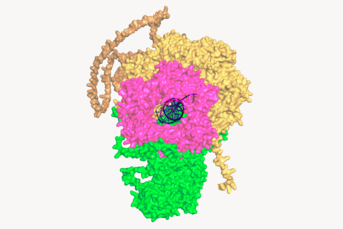
At Rockefeller, his research will investigate the fundamental gene expression process during which a series of molecular machines act in concert to transcribe DNA code into RNA, and then translate RNA into protein. He is interested in interactions among the molecular machines responsible for the synthesis, translation, and degradation of messenger RNA in bacterial cells. Dr. Liu also plans to study how the DNA-transcribing enzyme known as RNA polymerase reads through nucleosomes, the DNA-organizing units found in eukaryotic cells, and how this process is regulated by additional factors and epigenetic modifications. In addition to single-molecule biophysical tools, Dr. Liu will leverage the power of modern biochemical and genomic approaches to elucidate the molecular mechanism of these complex processes.
“I believe Shixin’s particular approach of exploring the interplay between the motors responsible [for gene expression] will contribute a unique perspective to the basic biology of gene regulation, as well as uncover implications for health and disease,” says Dr. Tessier-Lavigne. “I anticipate seeing great things from him as his career develops.”
A native of China and the child of two biology teachers, Dr. Liu studied biology at the University of Science and Technology of China. His entry into the field coincided with a shift in the approaches taken by biologists. “I grew up in a period when biology really started to be understood in a quantitative or analytical manner, not just as a descriptive science,” he says. “I was drawn to the idea of applying approaches from physics, chemistry, or mathematics to explore the fundamentals of biology. Work at the interface between these diverse disciplines will naturally bring together people with varied expertise and spark novel approaches to uncovering the secrets of life.”
Thomas Walz
Thomas Walz, a structural biologist who uses cutting-edge electron microscopy techniques to better our understanding of processes involving biological membranes, will join Rockefeller’s faculty as a tenured professor on September 1. As head of the Laboratory of Molecular Electron Microscopy, Dr. Walz will take advantage of the university’s recently acquired cryo-electron microscopes, which can capture molecular structures in unprecedented detail, to advance his research on macromolecular complexes and proteins embedded in cellular membranes.
“Tom has unparalleled expertise using sensitive new techniques to explore the architecture of biologically important molecules, and as a result, their function,” says Dr. Tessier-Lavigne. “His addition to our faculty will further strengthen our thriving team of structural biologists, and his deep knowledge of modern electron microscopy tools will make him an indispensable colleague for many within our community, helping us make the most of our recent investments in this technology.”
Dr. Walz began his scientific career studying the structures of proteins embedded in the membrane that surrounds cells, which is composed of two layers of molecules known as lipids. As a Ph.D. student at the University of Basel’s Biozentrum in Switzerland, he began work on the structure of aquaporin-1, a protein that forms a selective channel to allow water to travel in and out of cells. The structure resolved a long-standing riddle as to how aquaporins can efficiently conduct water while they are impermeable to protons, which should be able to move along with the water.
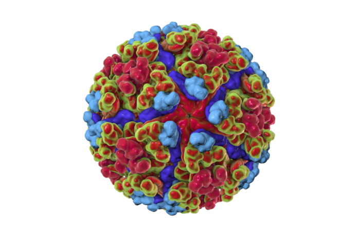
But long before that, the seed for his investigation of the unseen world was planted when, as a university student, he looked through an electron microscope at a drop of what appeared to be clean water but contained particles that looked like capsules designed for landing on the moon. These were viruses with their protein shells, called capsids.
“It was like looking into a different world. Some people are fascinated by the universe, I became fascinated by the very small structures that can’t be seen with the eye alone,” Dr. Walz says. “Because I am a very visual person, electron microscopy made sense for me, because you always see what you are studying.”
Following his Ph.D. work, Dr. Walz moved to the University of Sheffield in the United Kingdom, where as a postdoc he determined the two-dimensional structures of three photosynthetic complexes of membrane proteins from the bacterium Rhodobacter sphaeroides. After joining the faculty of Harvard Medical School in 1999, he continued studying the structures of other aquaporins, including aquaporin-0, a water channel in the eye’s lens that also acts as an adhesive for cell membranes, helping to form connections between cells.
“The images we obtained of aquaporin-0 had a high enough resolution that they also revealed the lipids in the surrounding membrane. That started to get me interested in how proteins interact with lipids and how proteins and lipids accommodate each other,” Dr. Walz says.
This interplay remains poorly understood. Proteins embedded in the cellular membrane are responsible for carrying out the membrane’s functions: relaying signals, allowing for cargo transport, catalyzing reactions, and mediating all interactions with the external environment and other cells. Studies continue to reveal the structures of these proteins, and as a result, how they carry out these activities. However, most of this structural work has been conducted on isolated membrane proteins in solution, without the lipid bilayer that is the membrane protein’s native environment. Meanwhile, cellular membranes contain thousands of different lipids, and it is being increasingly recognized that this diversity affects most membrane processes as well as the membrane proteins themselves.
Taking advantage of new tools in electron microscopy, Dr. Walz investigates the structure and function of membrane proteins within the context of the lipid environment and of macromolecular complexes, such as those that guide the transport of cargo throughout the cell. In addition to the ever-more-sophisticated software used to calculate high-resolution structures from noisy images, one of the major developments he employs, the direct electron detector device camera, enables scientists to record images and movies with unprecedented contrast and to compensate for the inevitable movement of specimens that occurs at this scale. Dr. Walz also uses nanodiscs—small patches of lipid bilayer stabilized by a scaffolding protein—to study how lipids affect membrane protein structure and function. Because they recreate a membrane protein’s native environment, nanodiscs are a significant improvement upon the detergents traditionally used in electron microscopy.
Dr. Walz is already collaborating with a number of Rockefeller scientists including Roderick MacKinnon, Jue Chen, Seth Darst, Günter Blobel, Tarun Kapoor, and Sebastian Klinge. He will guide the structural biologists at Rockefeller to become independent in the use of cryo-electron microscopy, and he also looks forward to collaborating with biologists from other disciplines to address questions in their research fields that are best tackled by this approach.
“I am looking forward to joining Rockefeller’s thriving community of structural biologists. Both the state-of-the-art electron microscopy tools and the university’s small, collegial environment were crucial factors in my decision to move my lab,” Dr. Walz says.


